-
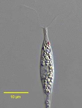
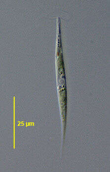
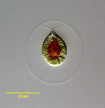
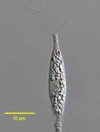
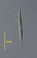
Hyalogonium klebsii (Klebs) Pascher,1927, a colorless volvocid flagellate with two equal anterior flagella. The body is elongate and fusiform. A red stigma is present on the left. Many small starch grains are seen in the cytoplasm.The nucleus is central. This genus may be confused with the euglenid Cyclidiopsis but Hyalogonium is smaller, biflagellate with thinner flagella, and lacks large paramylon bodies and the anterior canal opening. From standing rainwater pool near Boise, Idaho December 2005. DIC.
-
-
Hyalogonium klebsii (Klebs) Pascher,1927, a colorless volvocid flagellate with two equal anterior flagella. The body is elongate and fusiform. A red stigma is present on the left. Many small starch grains are seen in the cytoplasm.The nucleus is central. There are two anterior contractile vacuoles.This genus may be confused with the euglenid Cyclidiopsis but Hyalogonium is smaller, biflagellate with thinner flagella, and lacks large paramylon bodies and the anterior canal opening. From standing rainwater pool near Boise, Idaho December 2005. DIC.
-
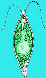
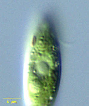
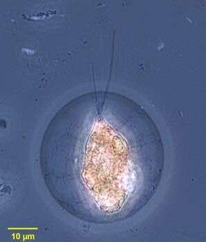
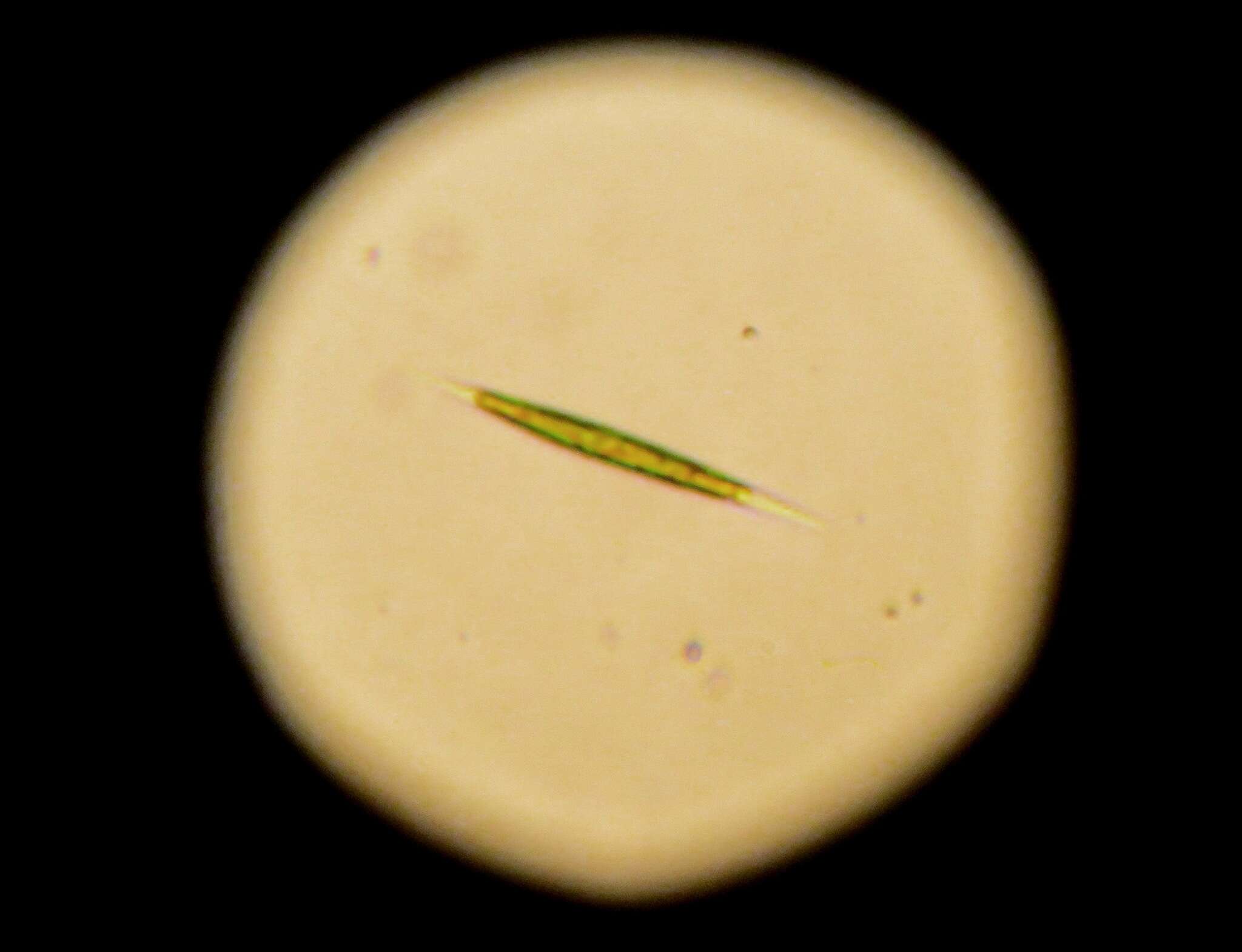
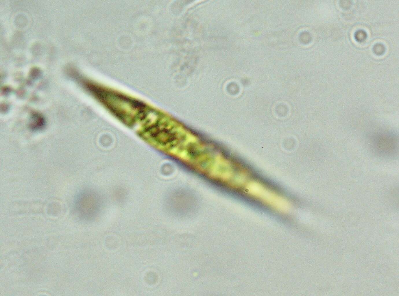
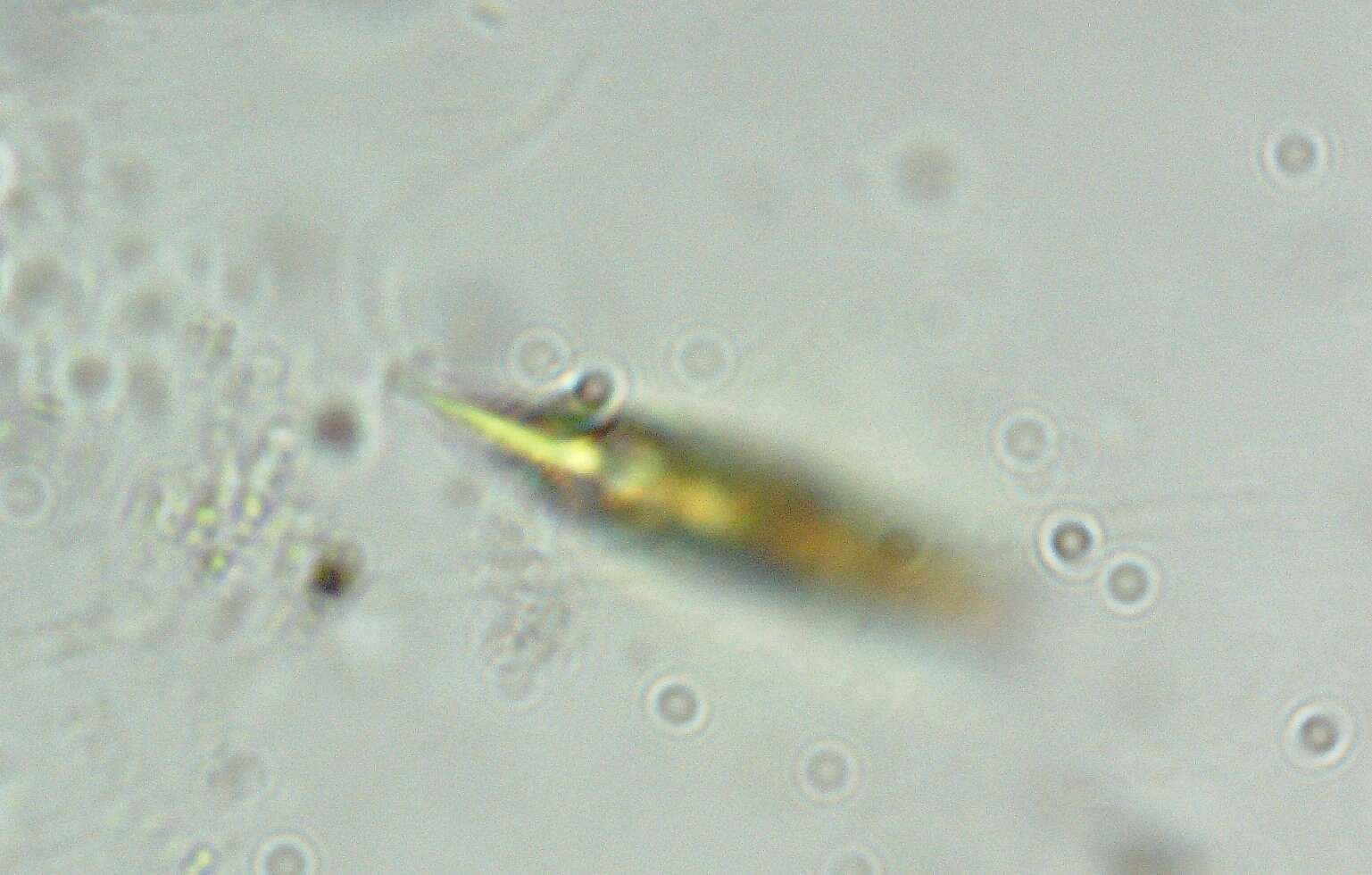
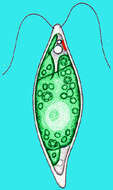
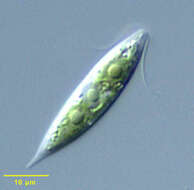
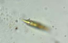
Chlorogonium, a small volvocid flagellate, with two equal anterior flagella. The cell body is fusiform. Chloroplasts are irregular. A stigma may be present. From standing rainwater pool near Boise, Idaho. Nomarski illumination.
-
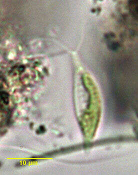
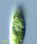
Chlorogonium, a volvocid green alga (Chlorophyta), the cell is spindle shaped with two flagella inserting at the anterior apex of the cell. The flagella beat with a breast-stroke like motion. This detail shows the anterior stigma or eyespot. Differential interference contrast optics.
-
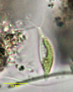
Chlorogonium, a volvocid green alga (Chlorophyta), the cell is spindle shaped with two flagella inserting at the anterior apex of the cell. The flagella beat with a breast-stroke like motion.
-
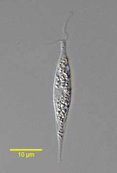
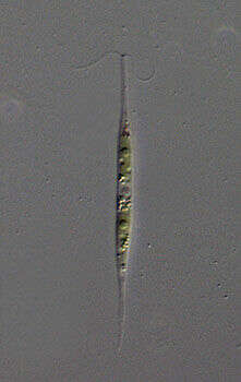
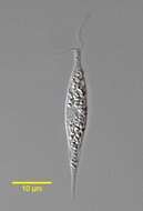
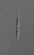
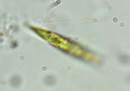
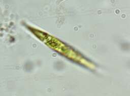
Portrait of the volvocid flagellate, Chlorogonium elongatum (Dangeard 1899). Collected from a freshwater pond near Boise, Idaho March 2005. DIC.
-
Portrait of the volvocid flagellate Chlorogonium elongatum (Dangeard 1899). Collected from a freshwater pond near Boise, Idaho March 2005. DIC
-
-
Phase contrast micrograph of living cell.
-
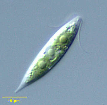
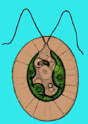
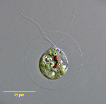
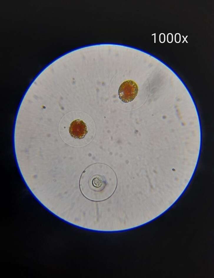
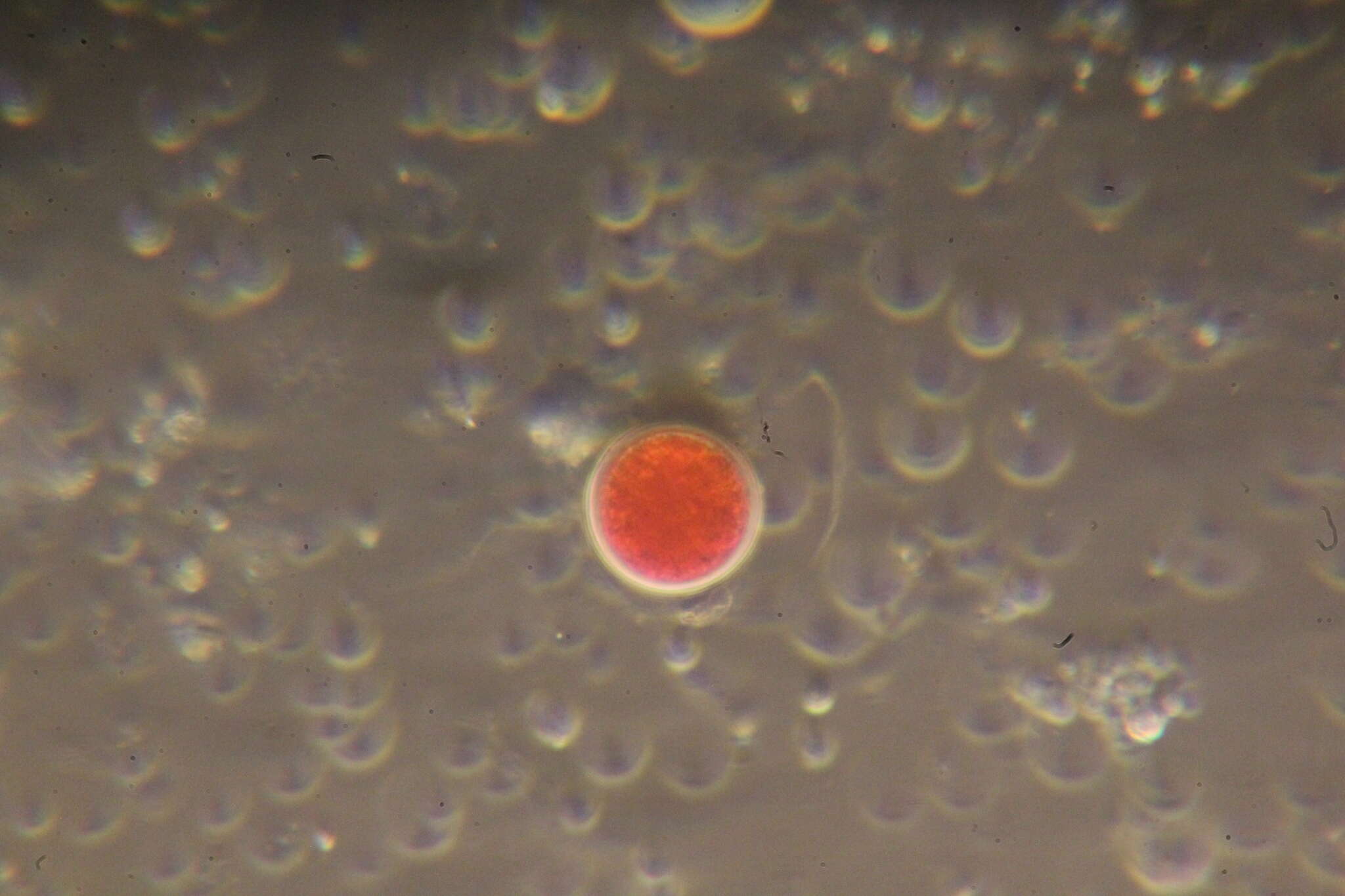
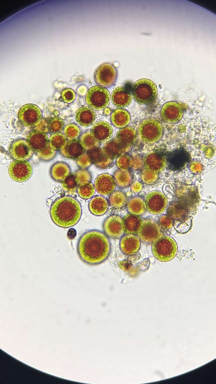
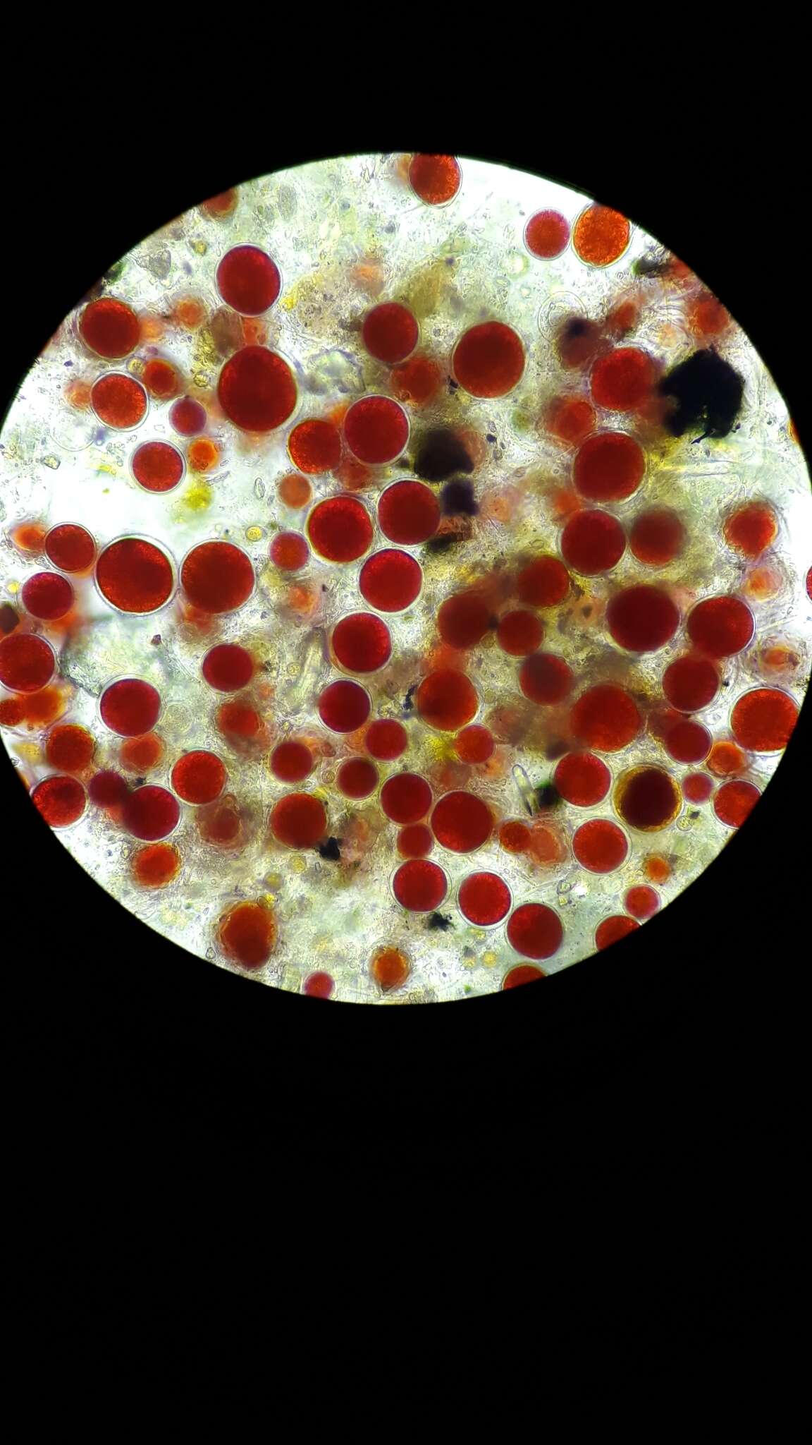
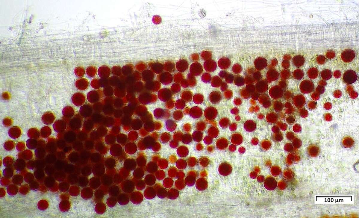
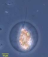
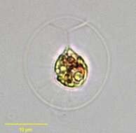
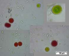
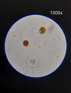
Portrait of Haematococcus pluvialis (Flotow, 1844), a widely distributed volvocid flagellate. Thin cytoplasmic strands traverse the clear mucilaginous layer to connect the protoplast to the spherical cell wall. Single large cup-shaped chloroplast. Two equal-length flagella are also seen traversing the mucilaginous layer. Reddish carotenoid pigments may obscure the stigma and chloroplast. From fish farm aquaculture pond near Boise, Idaho. Phase contrast.
-
Portrait of Haematococcus pluvialis (Flotow, 1844), a widely distributed volvocid flagellate. Thin cytoplasmic strands traverse the clear mucilaginous layer to connect the protoplast to the spherical cell wall. Single large cup-shaped chloroplast. Two equal-length flagella are also seen traversing the mucilaginous layer. Reddish carotenoid pigments may obscure the stigma and chloroplast. From fish farm aquaculture pond near Boise, Idaho. Brightfield.
-
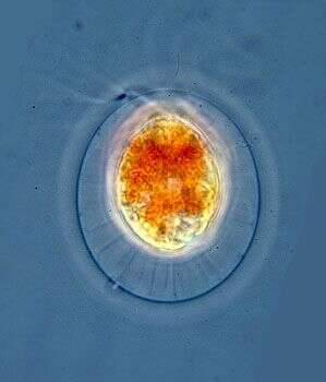
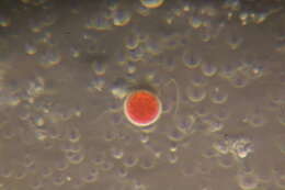
Portrait of Haematococcus pluvialis (Flotow, 1844), a widely distributed volvocid flagellate. Thin cytoplasmic strands traverse the clear mucilaginous layer to connect the protoplast to the spherical cell wall. Single large cup-shaped chloroplast. Two equal-length flagella are also seen traversing the mucilaginous layer. Reddish carotenoid pigments (concentrated in cell center here) may obscure the stigma and chloroplast. From ephemeral freshwater pool near Boise, Idaho, March, 2005. DIC.
-
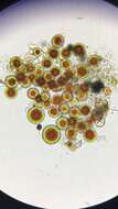
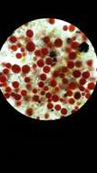
Portrait of Haematococcus pluvialis (Flotow, 1844), a widely distributed volvocid flagellate. Thin cytoplasmic strands traverse the clear mucilaginous layer to connect the protoplast to the spherical cell wall. Single large cup-shaped chloroplast. Two equal-length flagella are also seen traversing the mucilaginous layer. Reddish carotenoid pigments are concentrated in the cell center here. The inconspicuous stigma is seen at 1 o'clock near the proroplast surface. Several pyrenoids are visible here. There are multiple small contractile vacuoles. refractile cytoplasmic crystals are present in this individual. From ephemeral freshwater pool near Boise, Idaho, March, 2005. DIC.
-
-
-
-
-
-
-
-
-
-