portrait
Description:
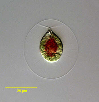
Portrait of Haematococcus pluvialis (Flotow, 1844), a widely distributed volvocid flagellate. Thin cytoplasmic strands traverse the clear mucilaginous layer to connect the protoplast to the spherical cell wall. Single large cup-shaped chloroplast. Two equal-length flagella are also seen traversing the mucilaginous layer. Reddish carotenoid pigments (concentrated in cell center here) may obscure the stigma and chloroplast. From ephemeral freshwater pool near Boise, Idaho, March, 2005. DIC.
Included On The Following Pages:
- Life (creatures)
- Cellular (cellular organisms)
- Eukaryota (eukaryotes)
- Archaeplastida (plants)
- Chloroplastida (green plants)
- Chlorophyta (chlorophytes)
- Chlorophyceae
- Chlamydomonadales
- Haematococcaceae
- Haematococcus
- Haematococcus lacustris
This image is not featured in any collections.
Source Information
- license
- cc-by-nc
- author
- William Bourland
- provider
- micro*scope
- original
- original media file
- visit source
- partner site
- micro*scope
- ID