-
-
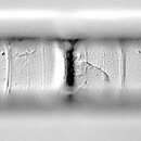
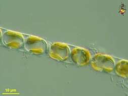
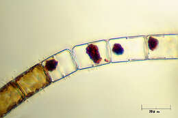
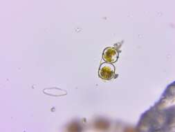
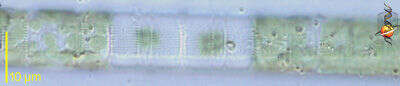
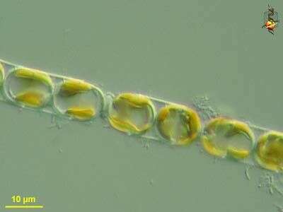
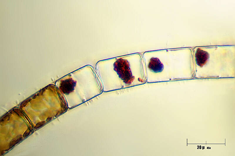
Melosira (mell-o-sigh-ra) is a centric diatom. The cells are like old-style hat boxes, or old-style pill boxes, or like petri-dishes. In Melosira, many cells are joined end to end to create a filament. The less substantial rings on the lower image are where the two halves of the frustule are joined together by the girdle bands, the more visible connections are where two cells are joined together. Each cell has a radial symmetry. As with other diatoms, plastids have chlorophylls a and c and so have a yellow brown colour. The lower picture reveals the individual disc-shaped plastids. Phase contrast.
-
Melosira (mellow-sire-a) nummuloides, filament forming centric diatom, with multiple small plastids within the cell. Dark ground illumination. Leptosiropsis (leapt-owe-sire-op-sis) torulosa, green alga with organic wall that is produced in layers. Phase contrast microscopy.
data on this strain.
-
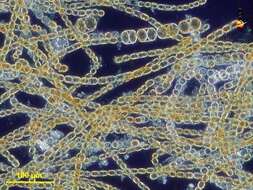
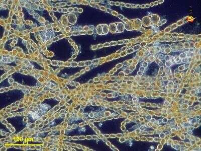
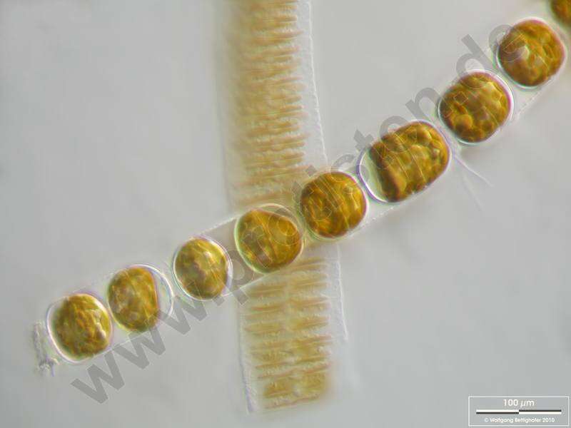
Colony with epibiotic bacteria chains. Scale bar indicates 100 µm. The image was built up using several photomicrographic frames with manual stacking technique. Sample from North Sea near Heligoland (spring diatom bloom). Images were taken using Zeiss Universal with Olympus C7070 CCD camera.
-
Torreblanca I, Catalunya, Espaa
-
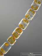
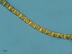
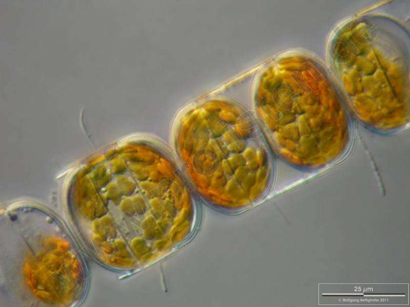
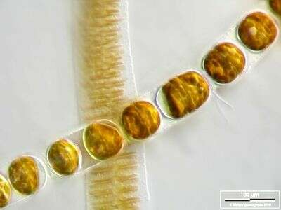
Melosira moniliformis.Colony with epibiotic bacteria chains. Scale bar indicates 25 m. The image was built up using several photomicrographic frames with manual stacking technique. Sample from North Sea near Heligoland (spring diatom bloom). Images were taken using Zeiss Universal with Olympus C7070 CCD camera.For more look at
www.protisten.de/english/gallery_main/gallery_main.htmlFor high-resolution images please ask postmaster@protisten.de.
-
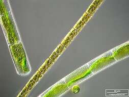
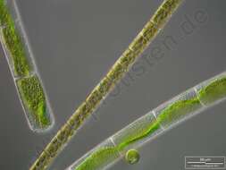
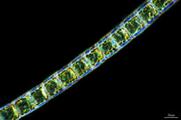
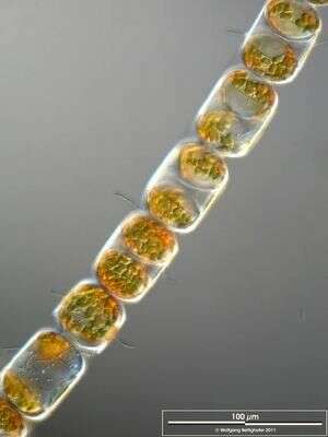
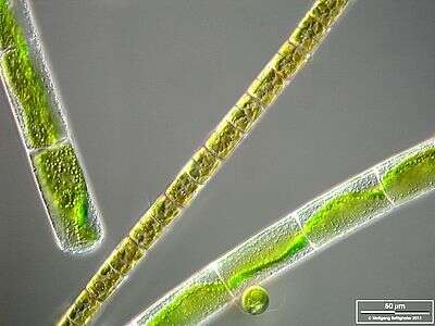
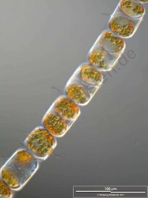
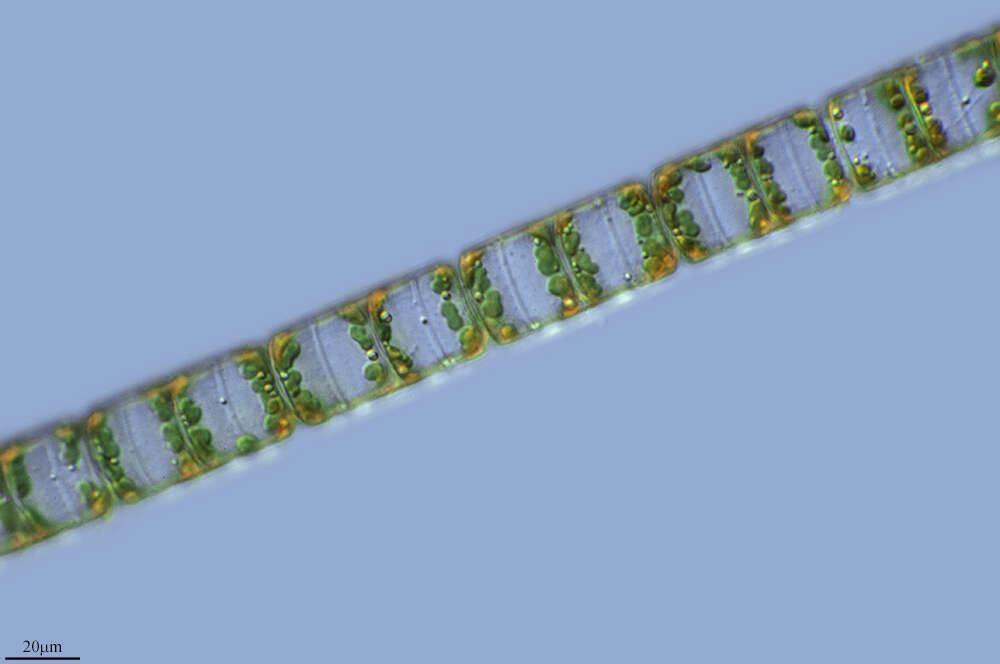
Melosira varians together with Mougeotia and Chlamydomonas. The scale bar indicates 50 µm. The specimen was gathered in the wetlands of Oderbruch (Oder valley 100 km north east of Berlin). The image was built up using several photomicrographic frames with manual stacking technique. Images were taken using Zeiss Universal with Olympus C7070 CCD camera.Image under Creative Commons License V 3.0 (CC BY-NC-SA).
-
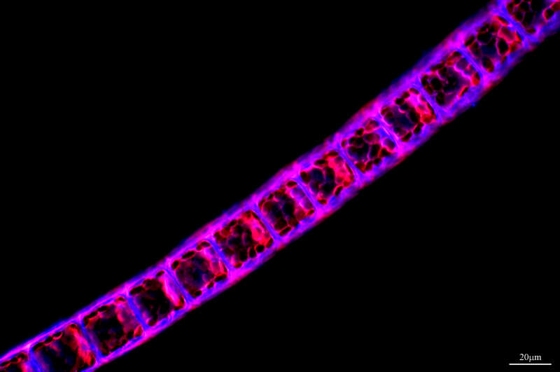
Melosira (mell-o-sire-a) is a centric diatom. The cells are like old-style hat boxes, or old-style pill boxes, or like petri-dishes. In Melosira, many cells are joined end to end to create a filament. The less substantial rings on the lower image are where the two halves of the frustule are joined together by the girdle bands, the more visible connections are where two cells are joined together. Each cell has a radial symmetry. As with other diatoms, plastids have chlorophylls a and c and so have a yellow brown colour. Differential interference contrast.
-
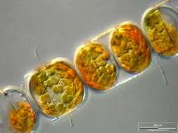
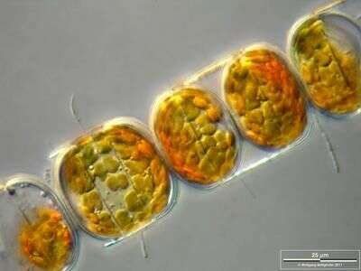
Melosira (mellow-sire-a) nummuloides, filament forming centric diatom, with multiple small plastids within the cell clearly shown in this micrograph. Differential interference microscopy.
data on this strain.
-
Colony with epibiotic bacteria chains. Scale bar indicates 25 µm. The image was built up using several photomicrographic frames with manual stacking technique. Sample from North Sea near Heligoland (spring diatom bloom). Images were taken using Zeiss Universal with Olympus C7070 CCD camera.
-
-
Melosira moniliformis.Colony with epibiotic bacteria chains. Scale bar indicates 100 m. The image was built up using several photomicrographic frames with manual stacking technique. Sample from North Sea near Heligoland (spring diatom bloom). Images were taken using Zeiss Universal with Olympus C7070 CCD camera.For more look at
www.protisten.de/english/gallery_main/gallery_main.htmlFor high-resolution images please ask postmaster@protisten.de.
-
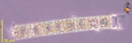
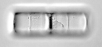
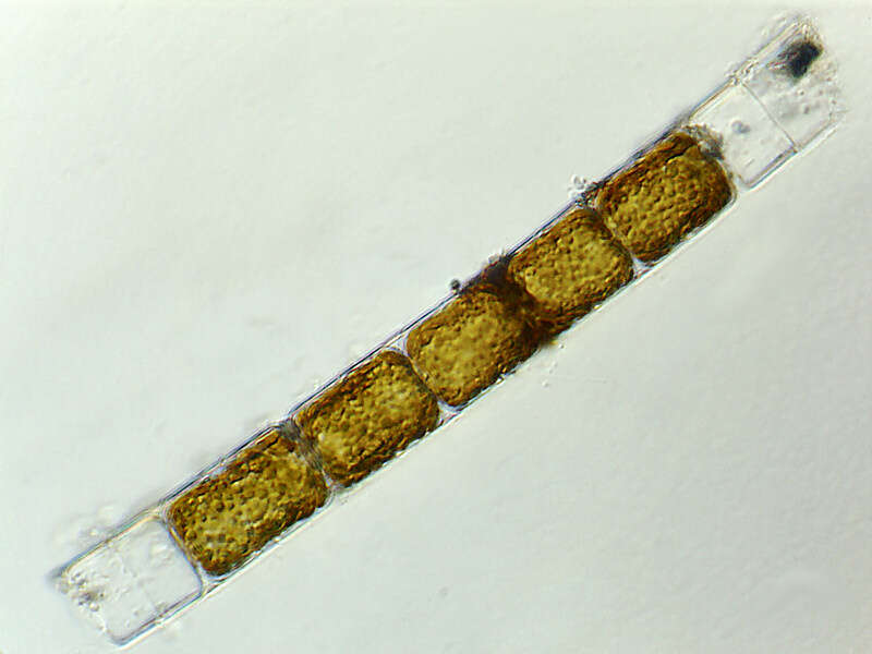
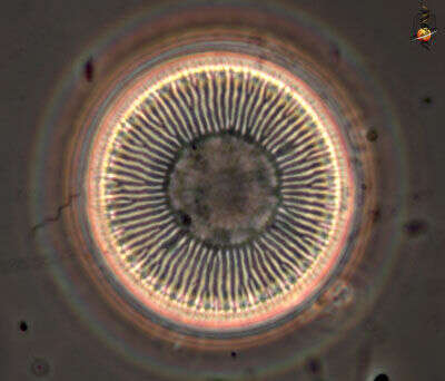
The surface of the siliceous valve of a Melosira cell. Phase contrast microscopy.
-
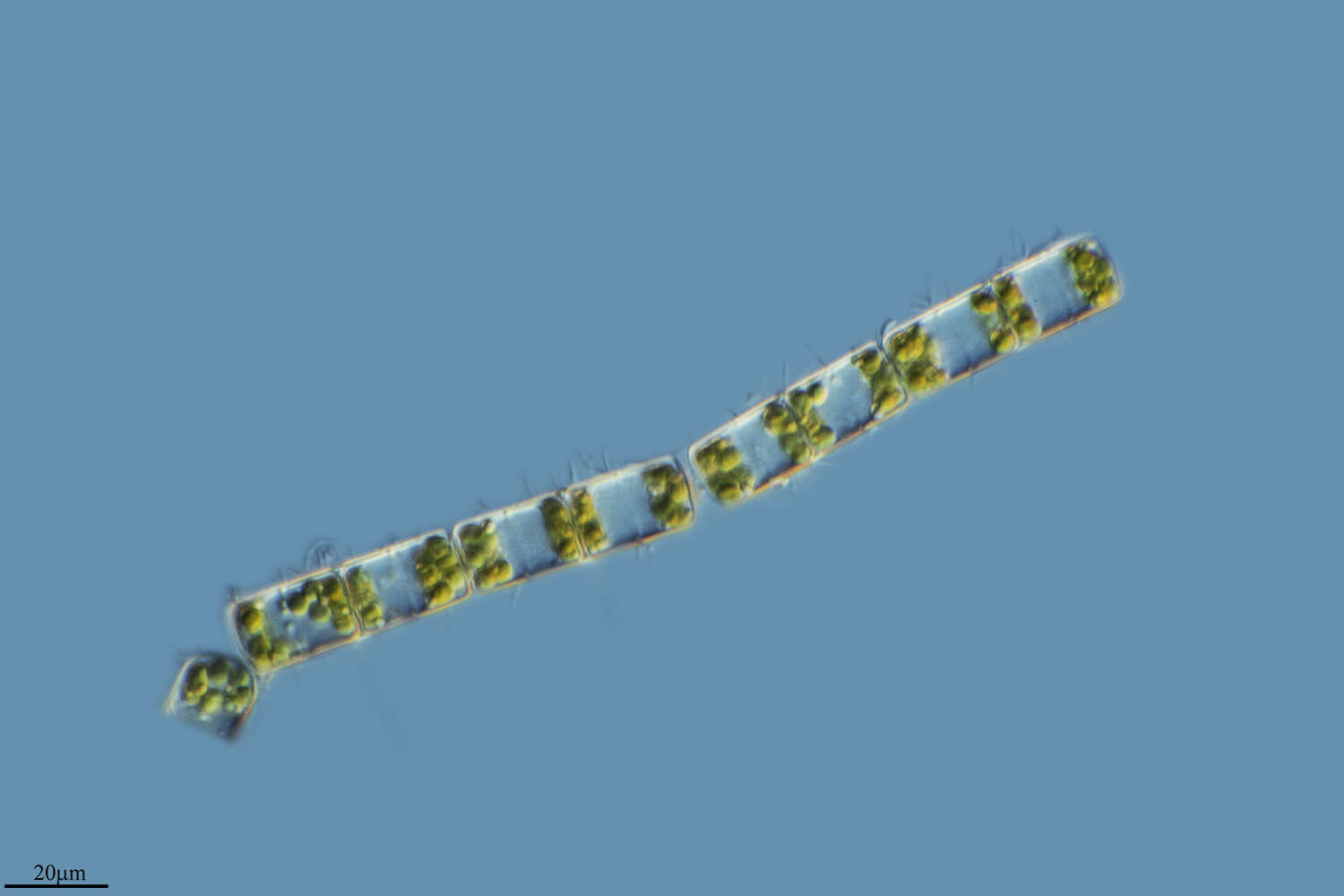
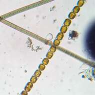
Melosira moniliformis accompanied by Fragilaria islandica. Scale bar indicates 100 µm. The image was built up using several photomicrographic frames with manual stacking technique. Sample from North Sea near Heligoland (spring diatom bloom). Images were taken using Zeiss Universal with Olympus C7070 CCD camera.
-
Melosira varians together with Mougeotia and Chlamydomonas. The scale bar indicates 50 m. The specimen was gathered in the wetlands of Oderbruch (Oder valley 100 km north east of Berlin). The image was built up using several photomicrographic frames with manual stacking technique. Images were taken using Zeiss Universal with Olympus C7070 CCD camera.For more look at
www.protisten.de/english/gallery_main/gallery_main.htmlFor high-resolution images please ask postmaster@protisten.de..
-
Melosira moniliformis accompanied by Fragilaria islandica. Scale bar indicates 100 m. The image was built up using several photomicrographic frames with manual stacking technique. Sample from North Sea near Heligoland (spring diatom bloom). Images were taken using Zeiss Universal with Olympus C7070 CCD camera.For more look at
www.protisten.de/english/gallery_main/gallery_main.htmlFor high-resolution images please ask postmaster@protisten.de.
-
Grvalos, La Rioja, Espaa
-
-
Talamanca, Catalonia, Spain
-
Sogo, Castille and Leon, Spain
-
Aguilar Del Rio Alhama, La Rioja, Spain
-
Aguilar del Ro Alhama, La Rioja, Espaa
-
-