-
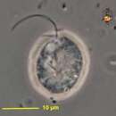
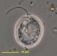
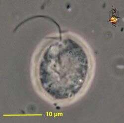
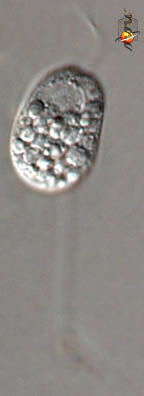
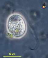
Allas (a-lass) rarely reported gliding flagellate from soils, rounded body, two flagella. Nuclear region is the light area in the centre of the cell. Contractile vacuole (usually there are two) near the point of flagellar insertion. Phase contrast.
-
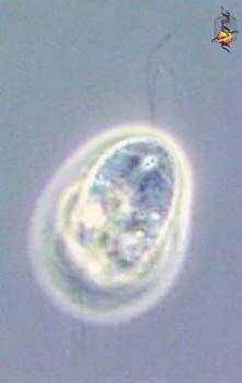
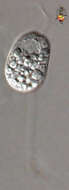
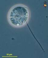
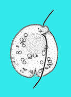
Protaspis (pro-tass-piss) A very common but little studied genus of gliding flagellates, two flagella inserted one in front of the other in a shallow ventral depression near the front of the cell. One flagellum trails behind the cell, one sweeps in front of the cell. There are caps (dictyosomes?) over the nucleus and these can be seen as two lines leading away from the site of flagellar insertion. Protaspis can produce pseudopodia and may eat diatoms. This individual has starchy inclusions. Phase contrast.
-
ATCC culture 50636.
-
Allas (a-lass), rarely reported gliding flagellate from soils, dividing. Division in most flagellates begins with a doubling of the n umber of flagella, and then a division furrow splits the cell longitudinally. Contractile vacuoles (usually there are two per cell) are the clear inclusions near the point of flagellar insertion. Phase contrast.
-
Protaspis (pro-tass-piss) A very common but little studied genus of gliding flagellates, two flagella inserted one in front of the other in a shallow ventral depression near the front of the cell. One flagellum trails behind the cell, one sweeps in front of the cell. There are caps (dictyosomes?) over the nucleus and these can be seen as two lines leading away from the site of flagellar insertion. Protaspis can produce pseudopodia and may eat diatoms. This individual has starchy inclusions. Phase contrast.
-
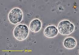
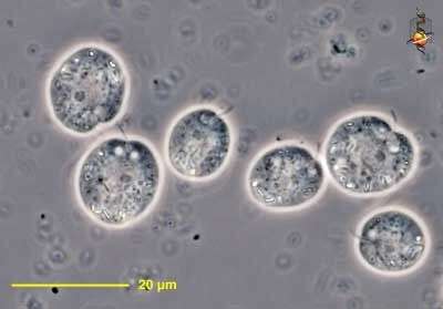
Allas (a-lass), rarely reported gliding flagellate from soils, rounded body, two flagella. Group of cells. Phase contrast.
-
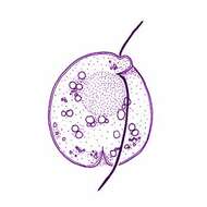
Protaspis (pro-tass-piss) A very common but little studied genus of gliding flagellates, two flagella inserted one in front of the other in a shallow ventral depression near the front of the cell. One flagellum trails behind the cell, one sweeps in front of the cell. Protaspis can produce pseudopodia and may eat diatoms. This individual has starchy inclusions. Phase contrast.
-
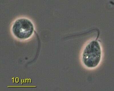
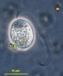
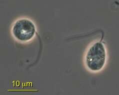
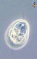
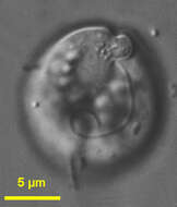
Protaspis (pro-tass-piss) is a medium-sized heterotrophic flagellate. Two flagella emerge close to each other from a point behind the apex of the cell and on the ventral side. The anterior flagellum is typically shorter than the posterior flagellum. The ventral side may give rise to pseudopodia which can enclose food - such as diatoms. Phase contrast microscopy.
-
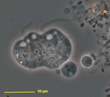
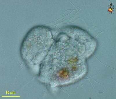
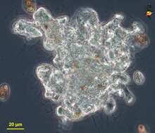
Protaspis (pro-tass-piss) is a medium-sized heterotrophic flagellate. Two flagella emerge close to each other from a point behind the apex of the cell and on the ventral side. The anterior flagellum is typically shorter than the posterior flagellum. The ventral side may give rise to pseudopodia which can enclose food - such as diatoms. In this image, many cells have fused into a syncitium. Phase contrast microscopy.
-
Protaspis (pro-tass-piss) is a medium-sized heterotrophic flagellate. Two flagella emerge close to each other from a point behind the apex of the cell and on the ventral side. The anterior flagellum is typically shorter than the posterior flagellum. The ventral side may give rise to pseudopodia which can enclose food - such as diatoms. In this image, many cells have fused into a syncitium. DIfferential interference microscopy.
-
Biflagellate protist with a ventral furrow and anterior depression from where the two flagella emerge. Species not identified. Isolated by M. Virginia Sanchez Puerta from Sippewissett Pond, Woods Hole, MA, USA. Photographed using DIC microscopy.
-
-
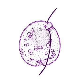
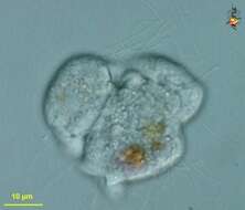
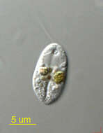
Protaspis (pro-tass-piss) obliqua Larsen and Patterson, 1990. Cells are slightly oval or roundish, 8 to 32 microns long, 10 to 27 microns wide, dorso-ventrally flattened and with thickened cortex. There is a ventral median groove, cell indented anteriorly and posteriorly where the groove meets margin. Subapically, the right margin of the groove forms a protrusion. With two flagella inserting under the protrusion, the anterior flagellum is about 0.5 times the length of the cell and the posterior flagellum is about 0.5 to 1.5 times the length of the cell. The nucleus is without nuclear caps, is located subapically in a median position, is rounded and is 5 to 13 microns in diameter. The cells may contain food particles or diatom up to 24 microns long. Commonly observed.
-
Protaspis obliqua Skuja, 1939. Cells are slightly oval or roundish, 8 to 32 microns long, 10 to 27 microns wide, dorso-ventrally flattened and with thickened cortex. There is a ventral median groove, cell indented anteriorly and posteriorly where the groove meets margin. Subapically, the right margin of the groove forms a protrusion. With two flagella inserting under the protrusion, the anterior flagellum is about 0.5 times the length of the cell and the posterior flagellum is about 0.5 to 1.5 times the length of the cell. The nucleus is without nuclear caps, is located subapically in a median position, is rounded and is 5 to 13 microns in diameter. The cells may contain food particles or diatom up to 24 microns long.
-
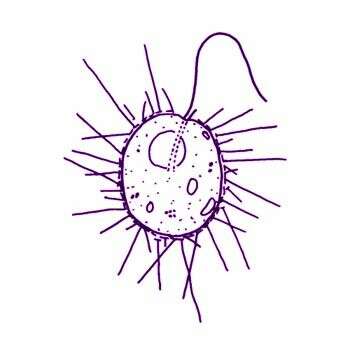
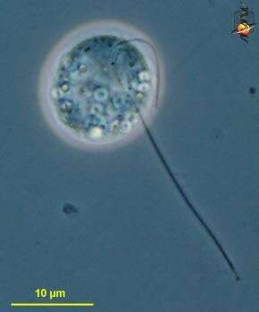
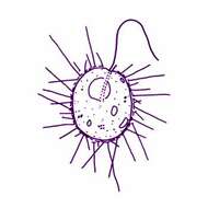
Thaumatomastix salina (Birch-Andersen) Beech and Moestrup, 1986. Cells are ovoid (7-12 microns x 8-15 microns), slightly compressed dorso-ventrally, and have a long flagellum 3/4-5/4 of the cell length. A short flagellum, which is rarely visible, emerges together with the long flagellum from what appears to be a very slight groove or depression located latero-anteriorly. A furrow-like structure is often noted running from the flagellar bases to the cell midline. Cells are solitary, and are most often observed attached to pieces of detritus. Cells occasionally move in a creeping motion, with the long flagellum trailing and gliding over the coverslip. In some cases cells swim freely with the long flagellum making irregular, arhythmical flicking motions. The cell cytoplasm has a granular appearance and is devoid of any kind of chloroplast, a diffuse area of a pale orange colour can often be noticed in the central part of the cell when phase contrast oil immersion optics are used. One cell was noted in an early stage of division where both flagella had replicated. Spine scales, varying in length, radiate from the entire cell surface. Flattened cells slough off their scales and scales of a second type, spineless body scales, can then be seen to be elliptical in outline.