Sobre
Educação
Discutir
Banco de Traços
Entrar
Criar conta
Idioma
Deutsch
English
Español
français
Nederlands
Piemontèis
Português do Brasil
suomi
Türkçe
Ελληνικά
македонски
Українська
العربية
简体中文
繁體中文
nomes no trilho de navegação
vernáculo
científico
Sobre
Educação
Discutir
Banco de Traços
Entrar
Criar conta
pt-BR
Deutsch
English
Español
français
Nederlands
Piemontèis
Português do Brasil
suomi
Türkçe
Ελληνικά
македонски
Українська
العربية
简体中文
繁體中文
nomes no trilho de navegação
vernáculo
científico
Life
»
…
»
Reino Animal
»
…
Life
»
Cellular
»
Eucariontes
»
Opisthokonta
»
Reino Animal
»
Bilateria
«
Xenacoelomorpha
coletar
início
dados
mídia
artigos
nomes
licença
qualquer licença
CC-BY
CC-BY-NC
CC-BY-NC-SA
CC-BY-SA
No copyright
tipo
qualquer tipo
imagem
mapa
som
vídeo
fornecedor
qualquer provedor
Arnold Arboretum Photo Gallery
Barcode of Life Data Systems
Barry Armstead Photography, ALA
BioImages, the virtual fieldguide, UK
Botanical Illustrations
Bugs for Bugs, Atlas of Living Australia
CalPhotos
CephBase
Eastfield College SEM Lab
eMammal
Femorale
Fishbase
Flickr BHL
Flickr Group
Harvard Museum of Comparative Zoology
iNaturalist
Moorea Biocode
Mycokeys
NMNH Collection
Phytokeys
PlanetScott
SailinSteve
SI Wild
SINA images
TreatmentBank
Turbellarian Taxonomic Database
USDA PLANTS images
Wikimedia Commons
Wolf Spiders of Australia, ALA
World Register of Marine Species
1
2
3
4
5
…
Last »
cc-by-nc-sa-3.0
confiável
cc-by-nc-sa-3.0
confiável
cc-by-nc-sa-3.0
confiável
cc-by-nc-sa-3.0
confiável
cc-by-nc-sa-3.0
confiável
cc-by-nc-sa-3.0
confiável
cc-by-sa-2.0
confiável
cc-by-nc-sa
confiável
cc-by-nc-sa-3.0
confiável
cc-by-nc-sa-3.0
confiável
cc-by-nc-sa-3.0
confiável
cc-by-nc-sa-3.0
confiável
cc-by-nc-sa-3.0
confiável
cc-by-sa-2.0
confiável
cc-by-nc-sa-3.0
confiável
cc-by-nc-sa-3.0
confiável
cc-by-nc-sa-3.0
confiável
cc-by-nc-sa-3.0
confiável
cc-by-nc-sa-3.0
confiável
cc-by-nc-sa-3.0
confiável
cc-by-nc-sa-3.0
confiável
cc-by-nc-sa-3.0
confiável
cc-by-nc-sa-3.0
confiável
cc-by-nc-sa-3.0
confiável
Imagem de Bursalia
cc-by-nc-sa-3.0
2010 Moorea Biocode
Moorea Biocode
All Biocode files are based on field identifications to the best of the researcher’s ability at the time.
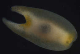
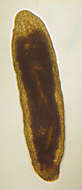
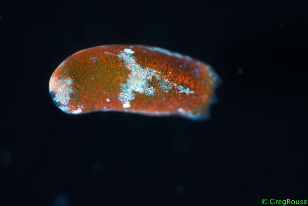
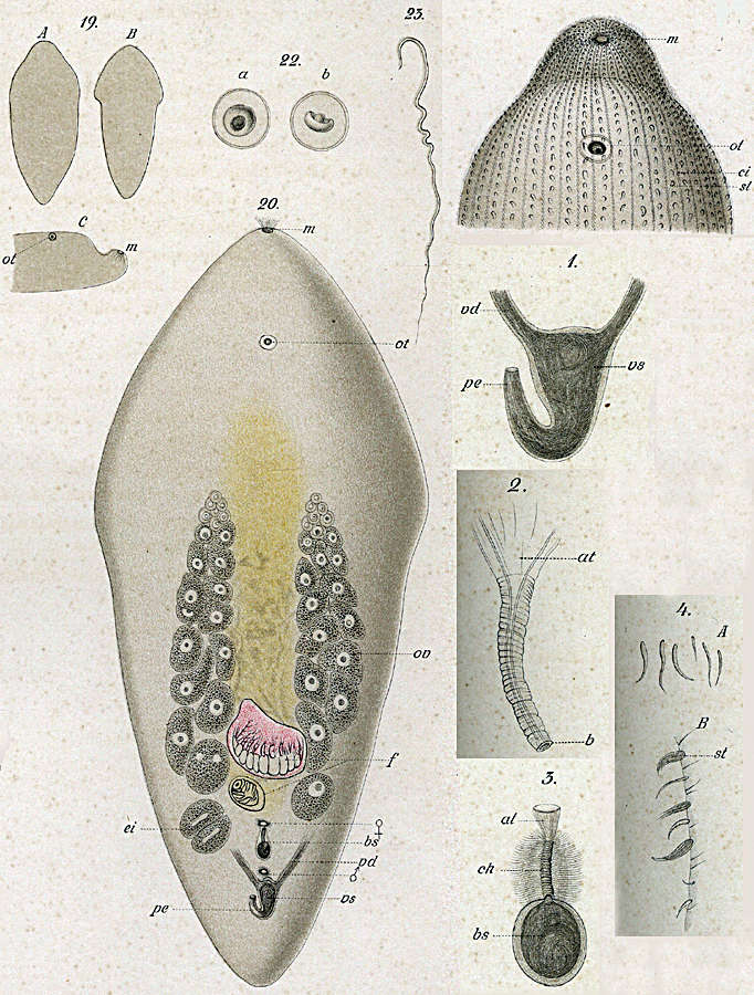
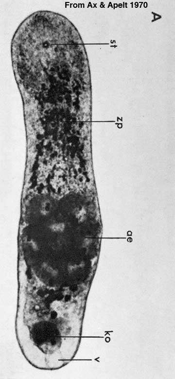
Imagem de Xenacoelomorpha
cc-by-nc-sa-3.0
2013 Moorea Biocode
Moorea Biocode
All Biocode files are based on field identifications to the best of the researcher’s ability at the time.
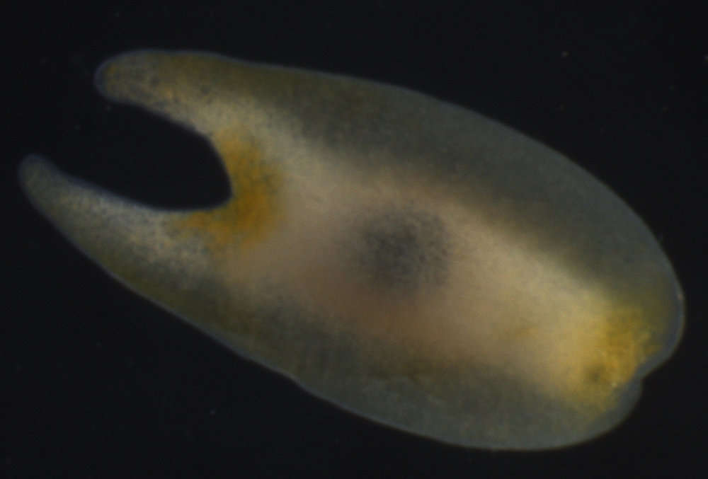
Imagem de Xenacoelomorpha
cc-by-nc-sa-3.0
National Science Foundation - Turbellarian Taxonomic Database
Turbellarian Taxonomic Database
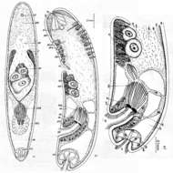
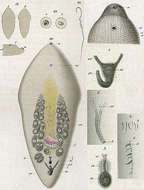
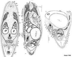
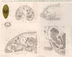
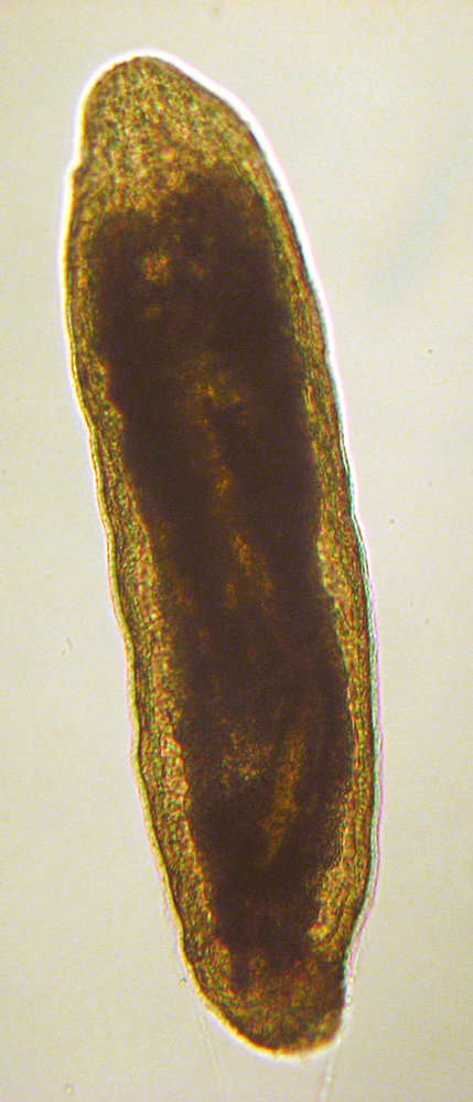
Imagem de Xenacoelomorpha
cc-by-nc-sa-3.0
National Science Foundation - Turbellarian Taxonomic Database
Turbellarian Taxonomic Database
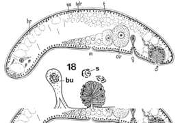
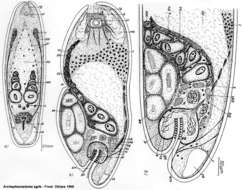
Imagem de Xenacoelomorpha
cc-by-nc-sa-3.0
National Science Foundation - Turbellarian Taxonomic Database
Turbellarian Taxonomic Database
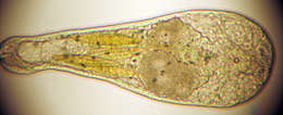
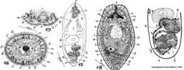
Imagem de Xenacoelomorpha
cc-by-nc-sa-3.0
National Science Foundation - Turbellarian Taxonomic Database
Turbellarian Taxonomic Database
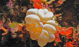
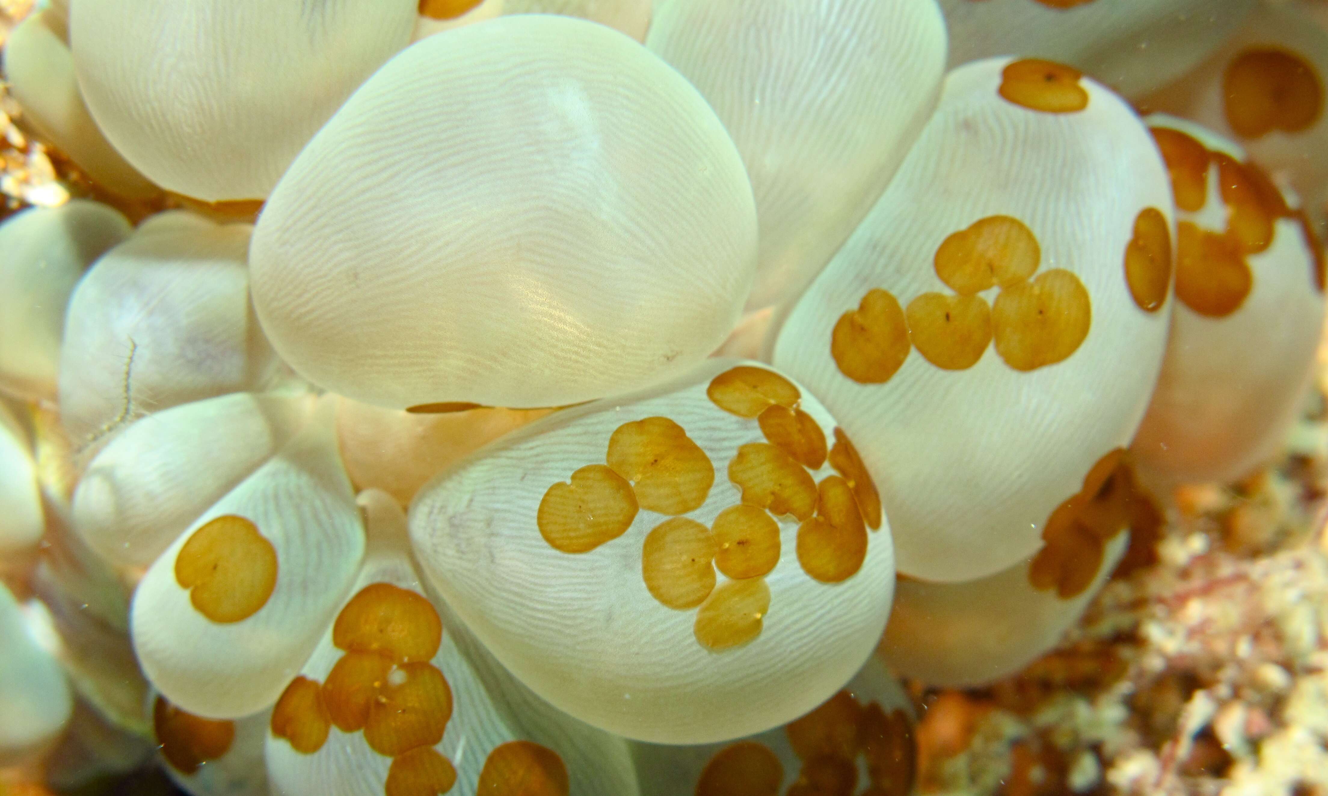
Acoelomorph Flatworms (Waminoa sp.) on Bubble Coral (Plerogyra sinuosa)
cc-by-sa-2.0
Bernard DUPONT
Flickr Group
Mabul, Sabah, Malaysia
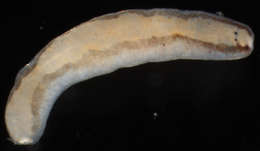
LAS MIRADAS DEL TERCER OJO DEL GUSANO DE ROSCOFF, SYMSAGITTIFERA ROSCOFFENSIS, PLAYA DE BALEA, RA DE AROUSA
cc-by-nc-sa
Proyecto Agua
Flickr Group
Valea, Galicia, Espaa
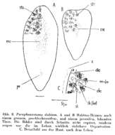
Imagem de Xenacoelomorpha
cc-by-nc-sa-3.0
2013 Moorea Biocode
Moorea Biocode
All Biocode files are based on field identifications to the best of the researcher’s ability at the time.
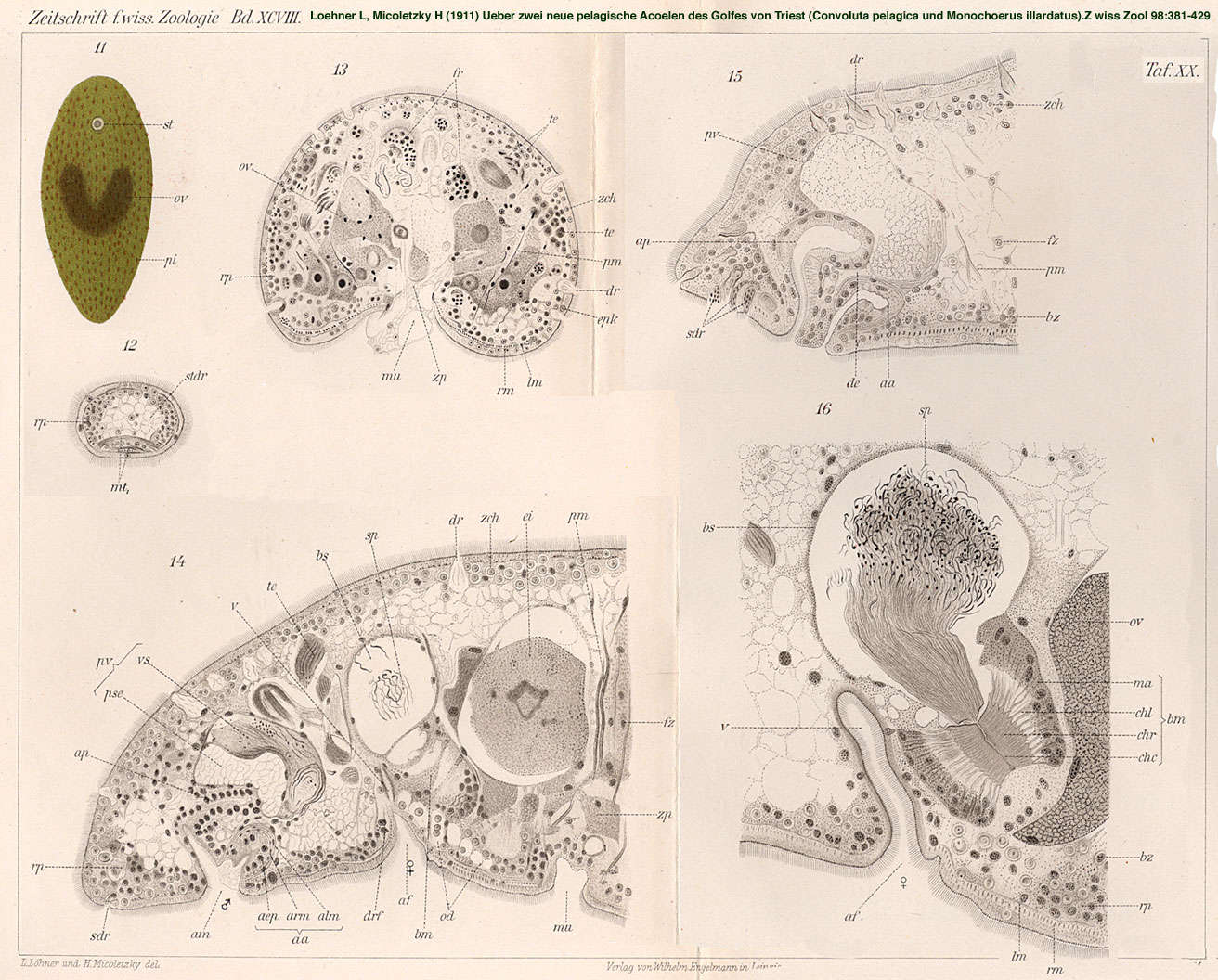
Imagem de Xenacoelomorpha
cc-by-nc-sa-3.0
National Science Foundation - Turbellarian Taxonomic Database
Turbellarian Taxonomic Database
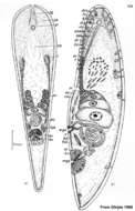
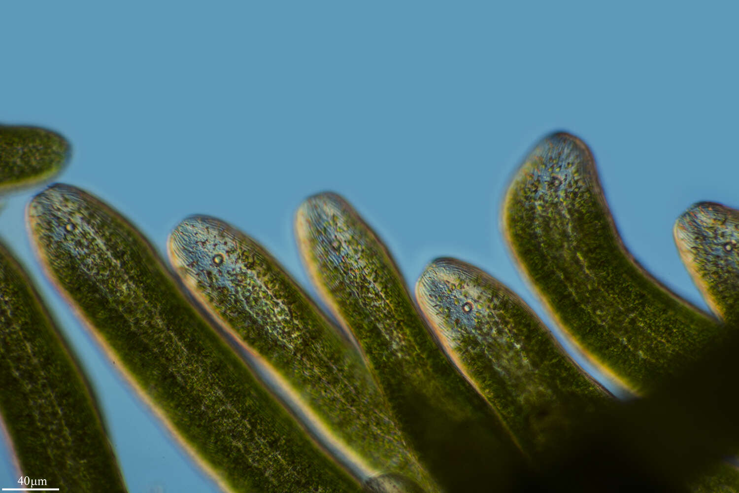
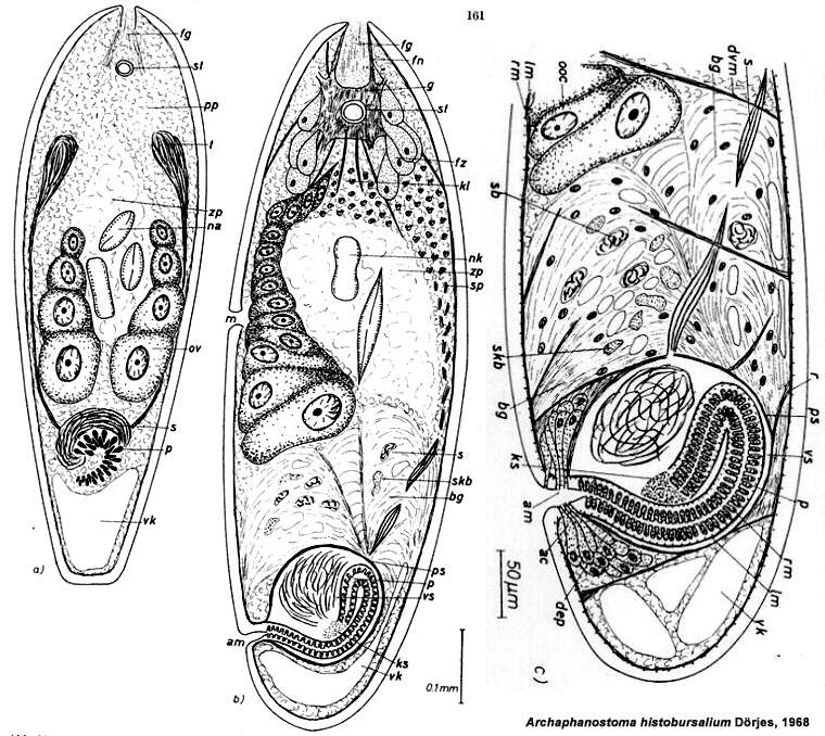
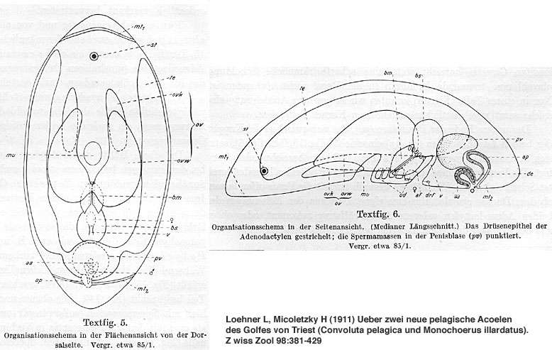
Imagem de Isodiametridae
cc-by-nc-sa-3.0
National Science Foundation - Turbellarian Taxonomic Database
Turbellarian Taxonomic Database
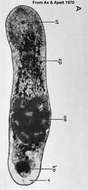
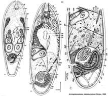
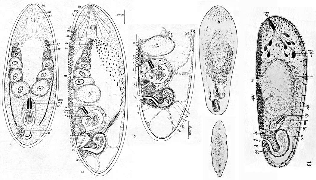
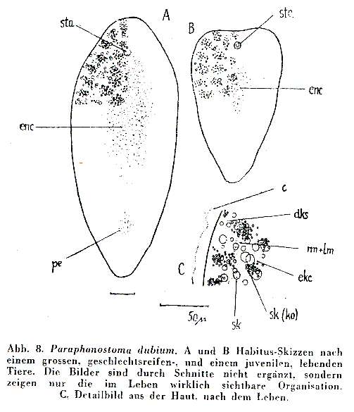
Imagem de Deuterogonaria
cc-by-nc-sa-3.0
National Science Foundation - Turbellarian Taxonomic Database
Turbellarian Taxonomic Database
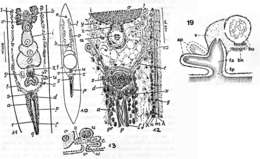
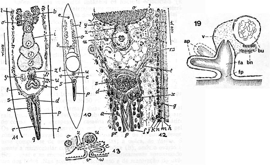
Imagem de Xenacoelomorpha
cc-by-nc-sa-3.0
National Science Foundation - Turbellarian Taxonomic Database
Turbellarian Taxonomic Database
Acoel Flatworms (Waminoa sp.) on Bubble Coral (Plerogyra sinuosa)
cc-by-sa-2.0
Bernard DUPONT
Flickr Group
Alungbanua, North Sulawesi, Indonesia
Imagem de Xenacoelomorpha
cc-by-nc-sa-3.0
2013 Moorea Biocode
Moorea Biocode
All Biocode files are based on field identifications to the best of the researcher’s ability at the time.
Imagem de Xenacoelomorpha
cc-by-nc-sa-3.0
National Science Foundation - Turbellarian Taxonomic Database
Turbellarian Taxonomic Database
Imagem de Isodiametridae
cc-by-nc-sa-3.0
National Science Foundation - Turbellarian Taxonomic Database
Turbellarian Taxonomic Database
Imagem de Aberrantospermata
cc-by-nc-sa-3.0
National Science Foundation - Turbellarian Taxonomic Database
Turbellarian Taxonomic Database
Imagem de Xenacoelomorpha
cc-by-nc-sa-3.0
National Science Foundation - Turbellarian Taxonomic Database
Turbellarian Taxonomic Database
Imagem de Xenacoelomorpha
cc-by-nc-sa-3.0
2013 Moorea Biocode
Moorea Biocode
All Biocode files are based on field identifications to the best of the researcher’s ability at the time.
Imagem de Xenacoelomorpha
cc-by-nc-sa-3.0
National Science Foundation - Turbellarian Taxonomic Database
Turbellarian Taxonomic Database
Imagem de Isodiametridae
cc-by-nc-sa-3.0
National Science Foundation - Turbellarian Taxonomic Database
Turbellarian Taxonomic Database
Imagem de Aberrantospermata
cc-by-nc-sa-3.0
National Science Foundation - Turbellarian Taxonomic Database
Turbellarian Taxonomic Database
Imagem de Xenacoelomorpha
cc-by-nc-sa-3.0
National Science Foundation - Turbellarian Taxonomic Database
Turbellarian Taxonomic Database
1
2
3
4
5
…
Last »