-
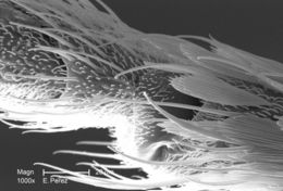
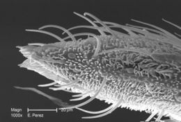
At a magnification of 1000X, twice that of PHIL 10557, this scanning electron micrograph (SEM) revealed some of the minute exoskeletal details found at the proboscis tip of an unidentified mosquito found deceased in the suburbs of Decatur, Georgia. The proboscis is the organ used by this, as well as other like insects, to feed upon the blood of a warm-blooded host, including human beings. What you see here, is the sheath that encases a pair of needle-sharp "stylets", which together are known as the "fascicle". The larger of the two stylets, known as the "labrum", when viewed in cross-section, takes on the shape of a "V", and acts as a gutter, which directs the ingested host blood towards the insect's mouth. The hair-like structures are known as "setae", and are really extensions of the insect's exoskeletal, chitinous covering. These setae act as sensory organs, transmitting impulses indicating changes in the organism's environment.Created: 2008
-
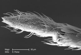
Magnified 500X, this scanning electron micrograph (SEM) revealed some of the minute exoskeletal details found at the proboscis tip of an unidentified mosquito found deceased in the suburbs of Decatur, Georgia. The proboscis is the organ used by this, as well as other like insects, to feed upon the blood of a warm-blooded host, including human beings. What you see here, is the sheath that encases a pair of needle-sharp "stylets", which together are known as the "fascicle". The larger of the two stylets, known as the "labrum", when viewed in cross-section, takes on the shape of a "V", and acts as a gutter, which directs the ingested host blood towards the insect's mouth. The hair-like structures are known as "setae", and are really extensions of the insect's exoskeletal, chitinous covering. These setae act as sensory organs, transmitting impulses indicating changes in the organism's environment.Created: 2008
-
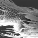
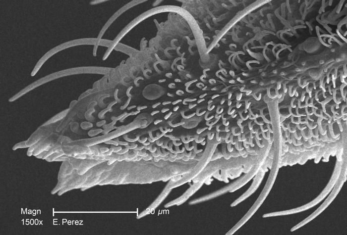
Magnified 1500X, this scanning electron micrograph (SEM) revealed some of the minute exoskeletal details found at the proboscis tip of an unidentified mosquito found deceased in the suburbs of Decatur, Georgia. The proboscis is the organ used by this, as well as other like insects, to feed upon the blood of a warm-blooded host, including human beings. What you see here, is the sheath that encases a pair of needle-sharp "stylets", which together are known as the "fascicle". The larger of the two stylets, known as the "labrum", when viewed in cross-section, takes on the shape of a "V", and acts as a gutter, directing the ingested host blood towards the insect's mouth. The hair-like structures are known as "setae", and are really extensions of the insect's exoskeletal, chitinous covering. These setae act as sensory organs, transmitting impulses indicating changes in the organism's environment.Created: 2008
-
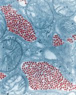
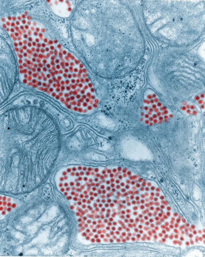
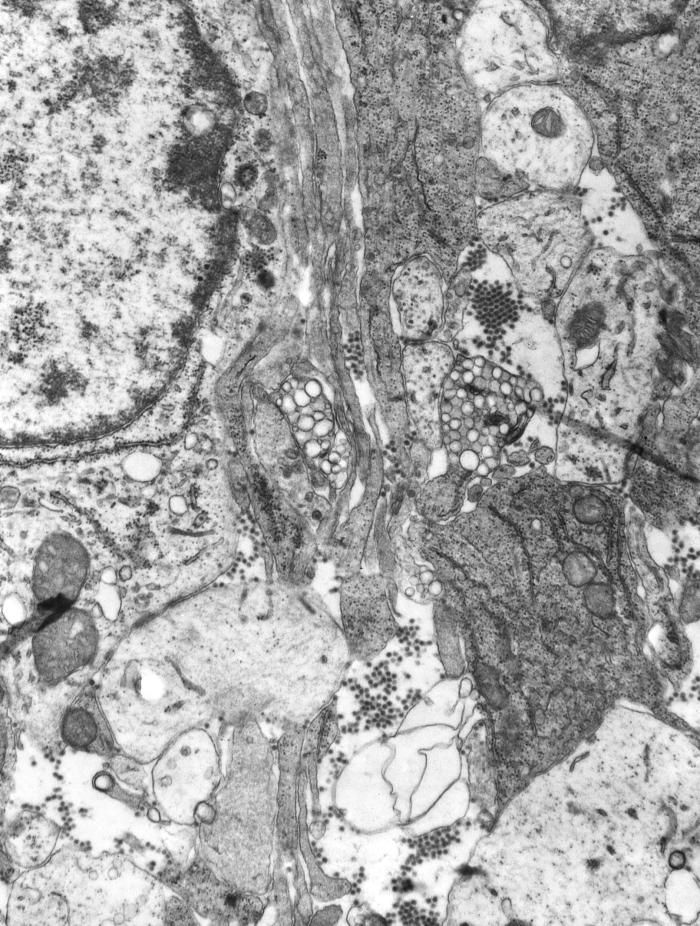
This colorized transmission electron micrograph (TEM) depicts a salivary gland that had been extracted from a mosquito, which was infected by the Eastern equine encephalitis (EEE) virus, which has been colorized red; magnified 83,900x.Created: 1968
-
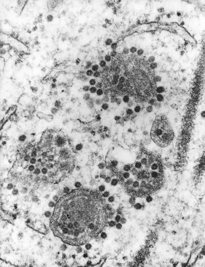
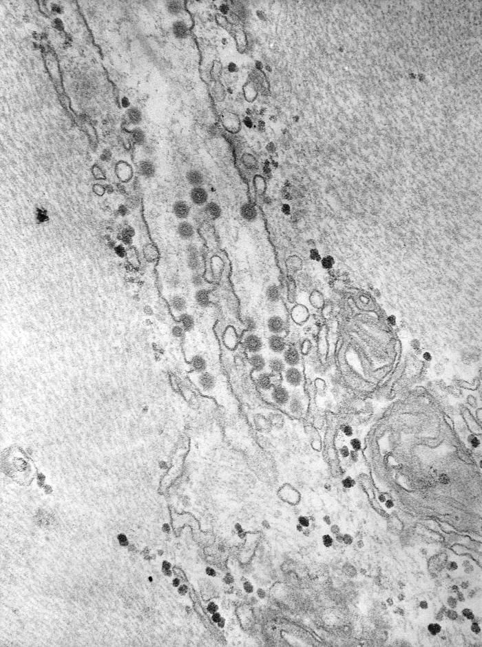
This 1975 transmission electron micrograph (TEM) revealed the presence of a number of Eastern Equine Encephalitis (EEE) virus virions that happened to be in a specimen of central nervous system tissue. EEE is an zoonotic arbovirus, which means that its spead to human beings through the bite of an infected mosquito. EEE virus (EEEV) occurs in the eastern half of the United States where it causes disease in humans, horses, and some bird species. Because of the high mortality rate, EEE is regarded as one of the most serious mosquito-borne diseases in the United States. EEE is a Togaviridae virus family member, and the genus Alphavirus.Created: 1975
-
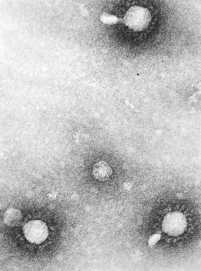
This negatively-stained 1975 transmission electron micrograph (TEM) revealed the presence of a number of Eastern equine encephalitis (EEE) virus virions in this tissue specimen. VEE is a Togaviridae family member, and a member of the genus Alphavirus.Eastern equine encephalitis (EEE) is a mosquito-borne viral disease. EEE virus (EEEV) occurs in the eastern half of the United States where it causes disease in humans, horses, and some bird species. Because of the high mortality rate, EEE is regarded as one of the most serious mosquito-borne diseases in the United States.Created: 1975
-
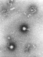
This negatively-stained 1975 transmission electron micrograph (TEM) revealed the presence of a number of Venezuelan equine encephalitis (VEE) virus virions in this tissue specimen, which had additionaly been fixed using phosphotungstic acid (PTA). This chemical is very electron-dense, and due to proposed electrostatic forces of attraction, clings to the capsid surface of each viral particle or virion, thereby, highlighting the presence of such pathogens. VEE is a Togaviridae family member, and a member of the genus Alphavirus.Created: 1975
-
This 1975 transmission electron micrograph (TEM) revealed the presence of a number of Eastern Equine Encephalitis (EEE) virus virions that happened to be in a specimen of central nervous system tissue. EEE is a zoonotic arbovirus, which means that its spead to human beings through the bite of an infected mosquito. EEE virus (EEEV) occurs in the eastern half of the United States where it causes disease in humans, horses, and some bird species. Because of the high mortality rate, EEE is regarded as one of the most serious mosquito-borne diseases in the United States. EEE is a Togaviridae virus family member, and the genus Alphavirus.Created: 1975
-
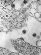
This negatively-stained transmission electron micrograph (TEM) revealed the presence of numerous Semliki Forest virus virions, which were present in a muscle tissue specimen. Named for the region in which they were isolate from mosquitoes, the Semliki Forest, Uganda, this virus is a Togaviridae family member, and the genus, Alphavirus.Created: 1975
-
This 1975 transmission electron micrograph (TEM) revealed the presence of a number of Eastern Equine Encephalitis (EEE) virus virions in this specimen of central nervous system tissue. EEE is an zoonotic arbovirus, which means that its spead to human beings through the bite of an infected arthropod, which in this case, is a mosquito. EEE virus (EEEV) occurs in the eastern half of the United States where it causes disease in humans, horses, and some bird species. Because of the high mortality rate, EEE is regarded as one of the most serious mosquito-borne diseases in the United States. EEE is a Togaviridae virus family member, and the genus Alphavirus.Created: 1975
-
This 1975 transmission electron micrograph (TEM) revealed the presence of a number of Eastern Equine Encephalitis (EEE) virus virions in this tissue specimen. EEE is an zoonotic arbovirus, which means that its spead to human beings through the bite of an infected arthropod, which in this case, is a mosquito. EEE virus (EEEV) occurs in the eastern half of the United States where it causes disease in humans, horses, and some bird species. Because of the high mortality rate, EEE is regarded as one of the most serious mosquito-borne diseases in the United States. EEE is a Togaviridae virus family member, and the genus Alphavirus.Created: 1975