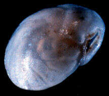
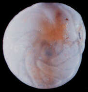
-
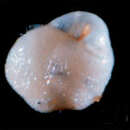
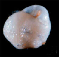
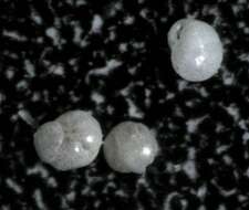
The aperture is at upper right. The test is 0.44 mm. across. Image courtesy of David B. Scott, Dalhousie University. This image was originally published in
Palaeologica Electronica, vol. 3, issue 2, and is used with the kind permission of that journal and the Paleontological Association.
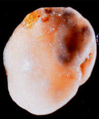
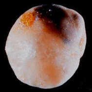
-
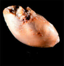
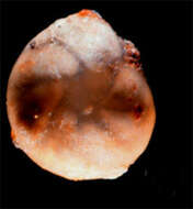
This test is broken at the aperture. Longest dimension is 1.53 mm. Image courtesy of David B. Scott, Dalhousie University. This image was originally published in
Palaeologica Electronica, vol. 3, issue 2, and is used with the kind permission of that journal and the Paleontological Association.
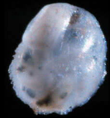
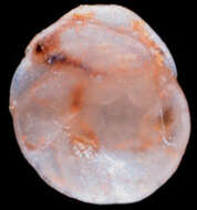
-
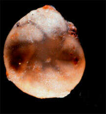
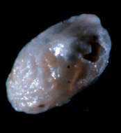
This photo is of the species holotype. The test is 1.5 mm across. Image courtesy of David B. Scott, Dalhousie University. This image was originally published in
Palaeologica Electronica, vol. 3, issue 2, and is used with the kind permission of that journal and the Paleontological Association.
-
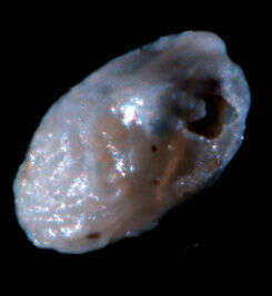
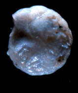
Image courtesy of David B. Scott, Dalhousie University. This image was originally published in
Palaeologica Electronica, vol. 3, issue 2, and is used with the kind permission of that journal and the Paleontological Association.
-
Test is 0.44 mm. across. Image courtesy of David B. Scott, Dalhousie University. This image was originally published in
Palaeologica Electronica, vol. 3, issue 2, and is used with the kind permission of that journal and the Paleontological Association.
-
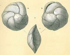
View of the holotype, showing the aperture. Image courtesy of David B. Scott, Dalhousie University. This image was originally published in
Palaeologica Electronica, vol. 3, issue 2, and is used with the kind permission of that journal and the Paleontological Association.
-
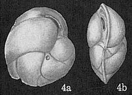
Image of the holotype. The test is 1.2 mm. across; aperture is at right. The chambers of the test are particularly clear in this photo. Image courtesy of David B. Scott, Dalhousie University. This image was originally published in
Palaeologica Electronica, vol. 3, issue 2, and is used with the kind permission of that journal and the Paleontological Association.
-
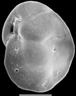
Image of the holotype. The aperture is at right. Image courtesy of David B. Scott, Dalhousie University. This image was originally published in
Palaeologica Electronica, vol. 3, issue 2, and is used with the kind permission of that journal and the Paleontological Association.
-
Image of the holotype. Test is 1.33 mm. across. Image courtesy of David B. Scott, Dalhousie University. This image was originally published in
Palaeologica Electronica, vol. 3, issue 2, and is used with the kind permission of that journal and the Paleontological Association.
-
Image of the holotype. Test is 1.33 mm. across. Image courtesy of David B. Scott, Dalhousie University. This image was originally published in
Palaeologica Electronica, vol. 3, issue 2, and is used with the kind permission of that journal and the Paleontological Association.
-
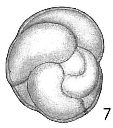
Image of the holotype; aperture is at top right, and the test is 0.67 mm. across. This subspecies is distinguished by the serrated edge on its test. Image courtesy of David B. Scott, Dalhousie University. This image was originally published in
Palaeologica Electronica, vol. 3, issue 2, and is used with the kind permission of that journal and the Paleontological Association.
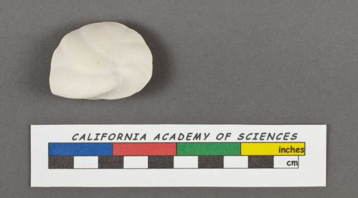
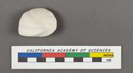
-
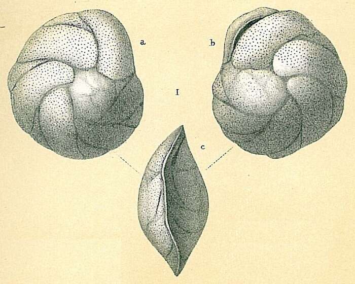
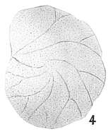
Credit: California Academy of Sciences Geology Orbigny d', A. D. (1826). Tableau méthodique de la classe des Céphalopodes. Annales des Sciences Naturelles. vol. 7: 96-169, 245-314., available online at (http://biodiversitylibrary.org/page/5753959) page(s): p. 282 n° 1 Model n° 41
-
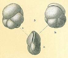
Credit: California Academy of Sciences Geology Orbigny d', A. D. (1826). Tableau méthodique de la classe des Céphalopodes. Annales des Sciences Naturelles. vol. 7: 96-169, 245-314., available online at (http://biodiversitylibrary.org/page/5753959) page(s): p. 282 n° 1 Model n° 41
-
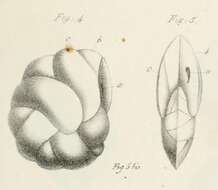
Orbigny d', A. D. (1826). Tableau méthodique de la classe des Céphalopodes. Annales des Sciences Naturelles. vol. 7: 96-169, 245-314., available online at (http://biodiversitylibrary.org/page/5753959) page(s): p. 282 n° 1 Model n° 41, pl. 15 fig. 4-5
-
Image source: Cushman, J.A. 1922. The Foraminifera of the Atlantic Ocean. Part 3. Textulariidae. Bull. U.S. Natl. Mus. 104.
-
Image source: Cushman, J.A. 1921. Foraminifera of the Philippine and adjacent seas. Bull. U.S. Natl. Mus. 100(4).
-
Image source: Goës, A. 1894. A Synopsis of the Arctic and Scandinavian recent marine Foraminifera hitherto discovered. Kong. Svenska Vetenskaps-Akademiens Handlingar 25(9): 1-127 + 25 pls. (Notae numerorum tenues mensuram indicant millimetricam).
-
Image source: Cushman, J.A. 1911. A Monograph of the Foraminifera of the North Pacific Ocean. Part II. Textulariidae. Bull. U.S. Nation. Mus 71: xiii+108 pp.
-
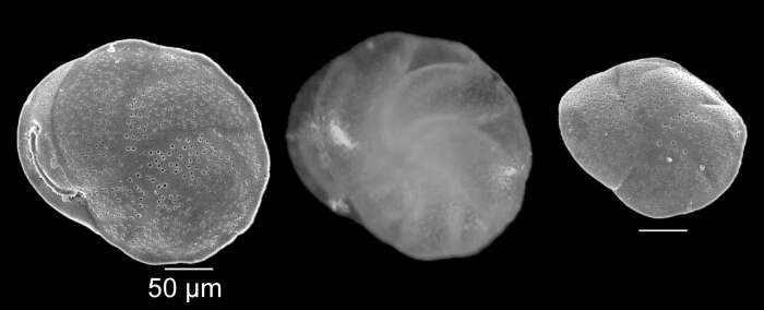
Scanning electron micrographs and photomicrographs of species from the northern Gulf of Cadiz continental shelf. Details about species distribution in: Mendes, I., Dias, J.A., Schönfeld, J., Ferreira, Ó., 2012. Distribution of living benthic foraminifera on the Northern Gulf of Cadiz continental shelf. Journal of Foraminiferal Research, 42(1), 18-38.
-
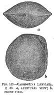
Cassidulina obtusa sensu Jones, R.W. 1994. The Challenger Foraminifera. Image source: Brady, H.B. (1884) Pl. 54
-
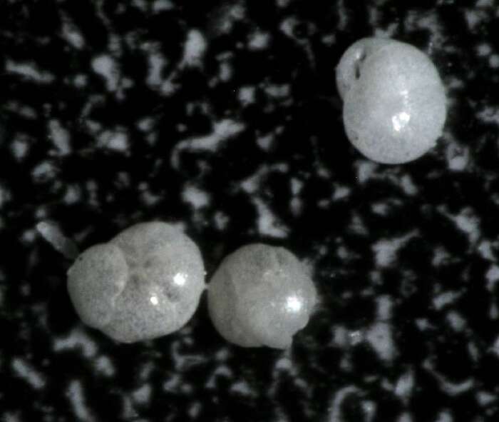
location: Hornsund Source: http://www.iopan.gda.pl/projects/biodaff/EMBS-06.html
-
Cassidulina teretis sensu Jones, R.W. 1994. The Challenger Foraminifera. Image source: Brady, H.B. (1884) Pl. 54
-
Image source: Todd, R. 1965. The Foraminifera of the Tropical Pacific Collections of the ”Albatross”, 1899-1900. Part 4. Rotaliform families and planktonic families [End of Volume]. Bull. U.S. Nation. Mus 161: v+139 pp.+28 pls.
-
Wanganui Bight, 57 m depth, Plate 8 in Hayward, B.W., Grenfell, H.R., Reid, C.M., Hayward, K.A. 1999. Recent New Zealand shallow-water benthic Foraminifera: Taxonomy, ecologic distribution, biogeography, and use in paleoenvironmental assessment. Institute of Geological and Nuclear Sciences Monograph 21, 258 p.