-
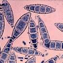
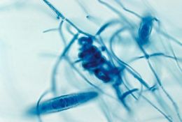
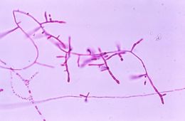
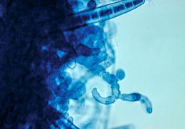
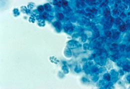
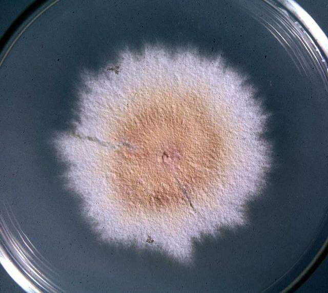
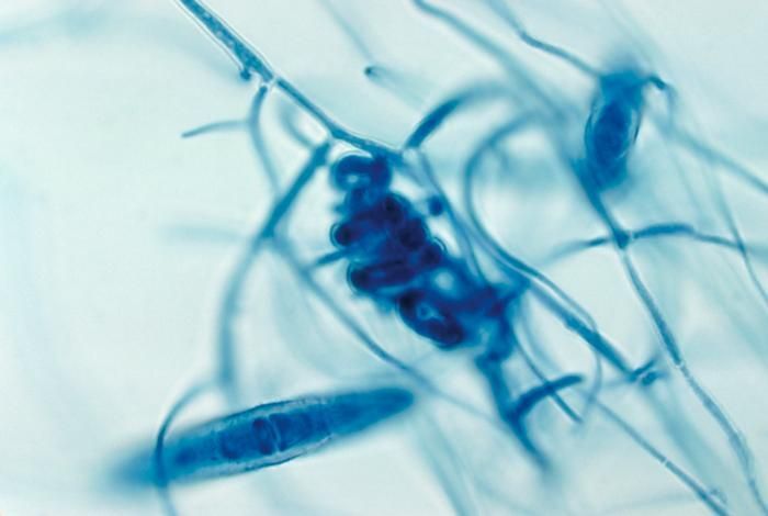
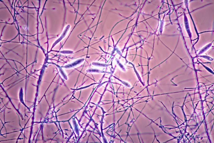
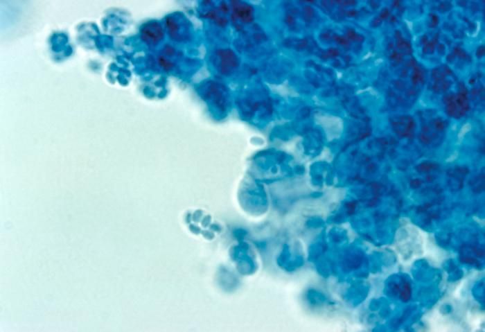
Mushroom Observer Image 55344: Microsporum gypseum (E. Bodin) Guiart & Grigoraki
-
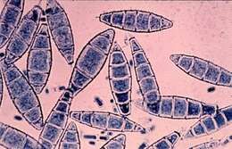
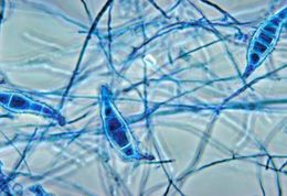
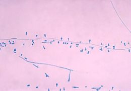
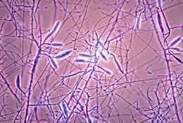
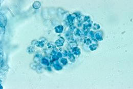
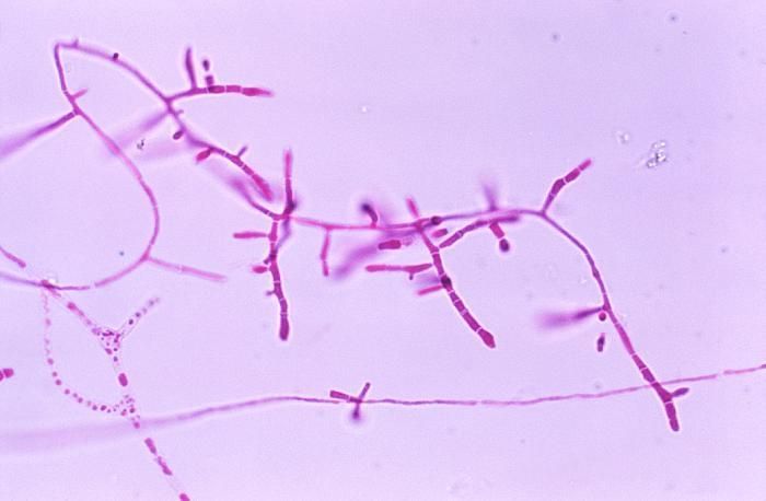
Mushroom Observer Image 55345: Microsporum gypseum (E. Bodin) Guiart & Grigoraki
-
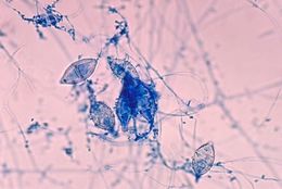
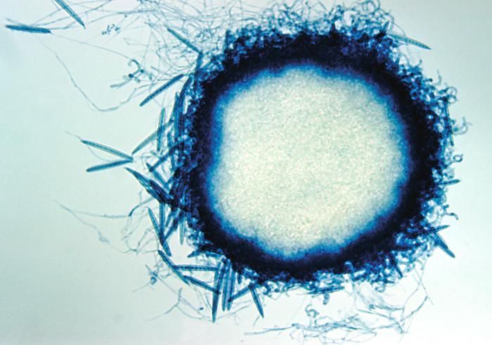
Mushroom Observer Image 55346: Microsporum gypseum (E. Bodin) Guiart & Grigoraki
-
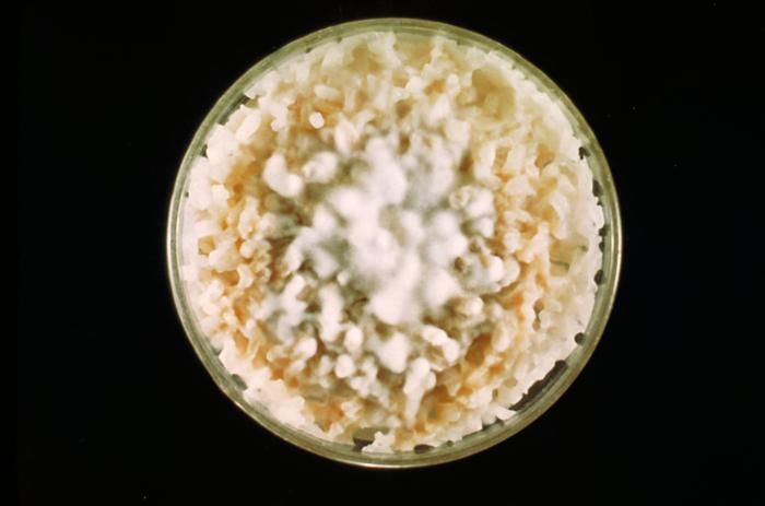
This image shows a culture of Microsporum canis fungi growing on boiled polished rice grains.Created: 1962
-
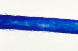
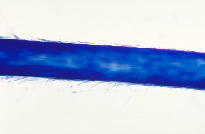
This slide was created during a hair perforation test for the zoophilic fungus Microsporum equinum.Created: 1978
-
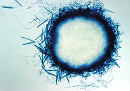
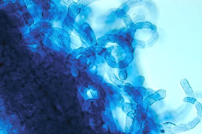
This photomicrograph shows the cleistothecium of the fungus Arthroderma grubyi, formerly Nannizzia grubyia.Created: 1961
-
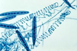
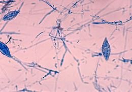
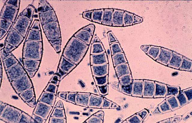
This micrograph depicts the irregularly shaped macroconidia from the fungal organism Microsporum distortum.Created: 1970
-
This photomicrograph shows the macroconidia of the zoophilic fungus Microsporum equinumCreated: 1978
-
This micrograph reveals spirals and macroconidia from the edge of an immature cleistothecium of the A. grubyi fungus.Created: 1961
-
Magnified under a low magnification of 40X, this photomicrograph depicts the microconidia of the fungus Trichophyton mariatii.Created: 1979
-
This photomicrograph shows the microconidia of the zoophilic fungus Microsporum equinum.Created: 1978
-
This photomicrograph shows a mature cleistothecium of the fungus Arthroderma grubyi, formerly Nannizzia grubyia.Created: 1961
-
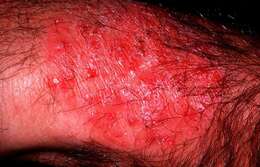
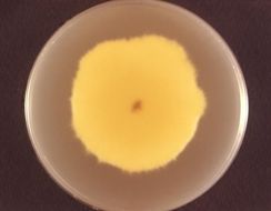
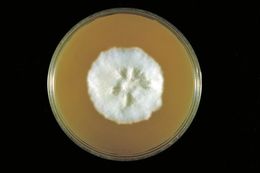
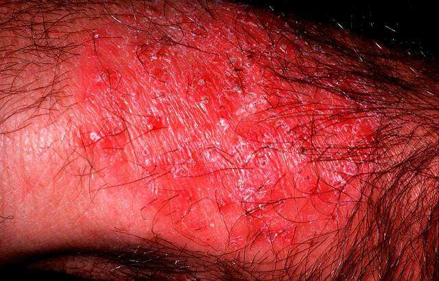
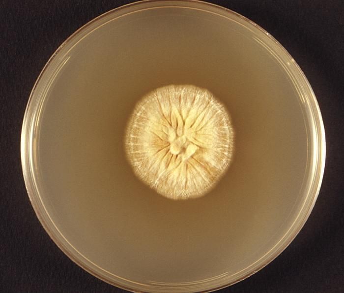
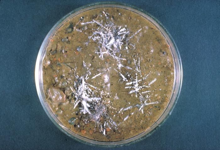
This photograph featured a Petri dish, which had been used to culture a colony of dermatophytic fungus, Microsporum ferrugineum.Dermatophytes are types of fungi that cause common skin, hair and nail infections. Infections caused by these fungi are also known by the names tinea and ringworm. It is important to emphasize that ringworm is not caused by a worm, but rather by a type of fungus called a dermatophyte. One example of a very common dermatophyte infection is athletes foot, which is also called tinea pedis. Another common dermatophyte infection affecting the groin area is jock itch, also known as tinea cruris.Created: 1974
-
This photomicrograph depicts the macroconidia of the zoophilic fungus Microsporum equinum.Created: 1978
-
This is the edge of a mature cleistothecium of the fungus Arthroderma grubyi, formerly Nannizzia grubyia.Created: 1961
-
This photomicrograph depicts the mycelia, conidiophores, and conidia of the fungus Microsporum gallinae.Created: 1978
-
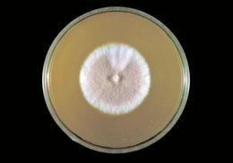
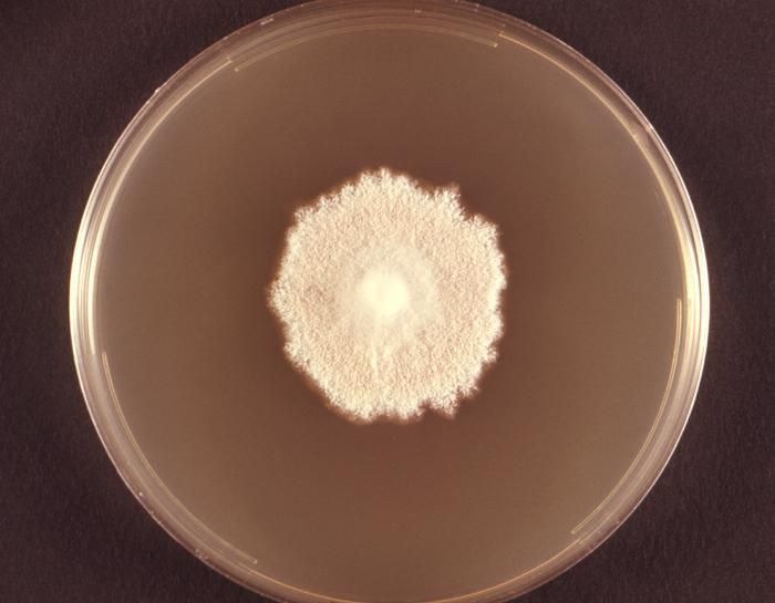
His hair-plate culture is growing the fungus Trichophyton terrestre.Created: 1963
-
This image shows the cleistothecium of the fungus Arthroderma grubyi, formerly known as Nannizzia grubyia.Created: 1961
-
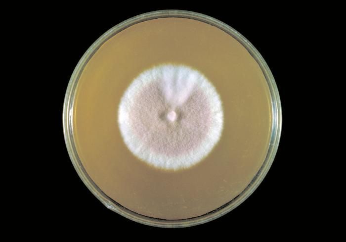
Viewed from the back, i.e., reverse, this image depicted a Petri dish containing Sabouraud's (SAB) dextrose agar, upon which a Microsporum persicolor fungal colony had been cultured. As seen in this reverse view, the colonial coloration can be yellow, or may even be a red-brown. From the front, as depicted in PHIL 10904 and 10906, the colonies can be white, or depending upon the Microsporum sp., may run the gamut, sporting a yellow, beige or cinnamon color, and display a flat, or glabrous, woolly or powdery texture.Created: 1973
-
This Sabourauds dextrose agar plate culture is growing T. terrestre fungus, rose-pigmented strain x231.Created: 1963
-
This photomicrograph shows the asci and ascospores of the fungus Arthroderma grubyi, formerly Nannizzia grubyia.Created: 1961
-
Photographed from the front, this image depicted a Petri dish containing cereal agar, upon which a Microsporum persicolor fungal colony had been cultured. As was the case here, the colonies can be white, or depending upon the Microsporum sp., may run the gamut, sporting a yellow, beige or cinnamon color, and display a flat, or glabrous, woolly or powdery texture. Taxonomically, M. persicolor is a member of the phylum Ascomycota. See PHIL 10905 for a reverse view of this colony, i.e., viewed from behind.Created: 1973
-
This Sabourauds dextrose agar plate culture is growing T. terrestre fungus, white strain x231, day 12.Created: 1963
-
This photomicrograph shows the asci and ascospores of the fungus Arthroderma grubyi, formerly Nannizzia grubyia.Created: 1961