-
Jaime Gonzalez-Cueto, Sigmer Quiroga, Jon Norenburg
Zookeys
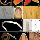
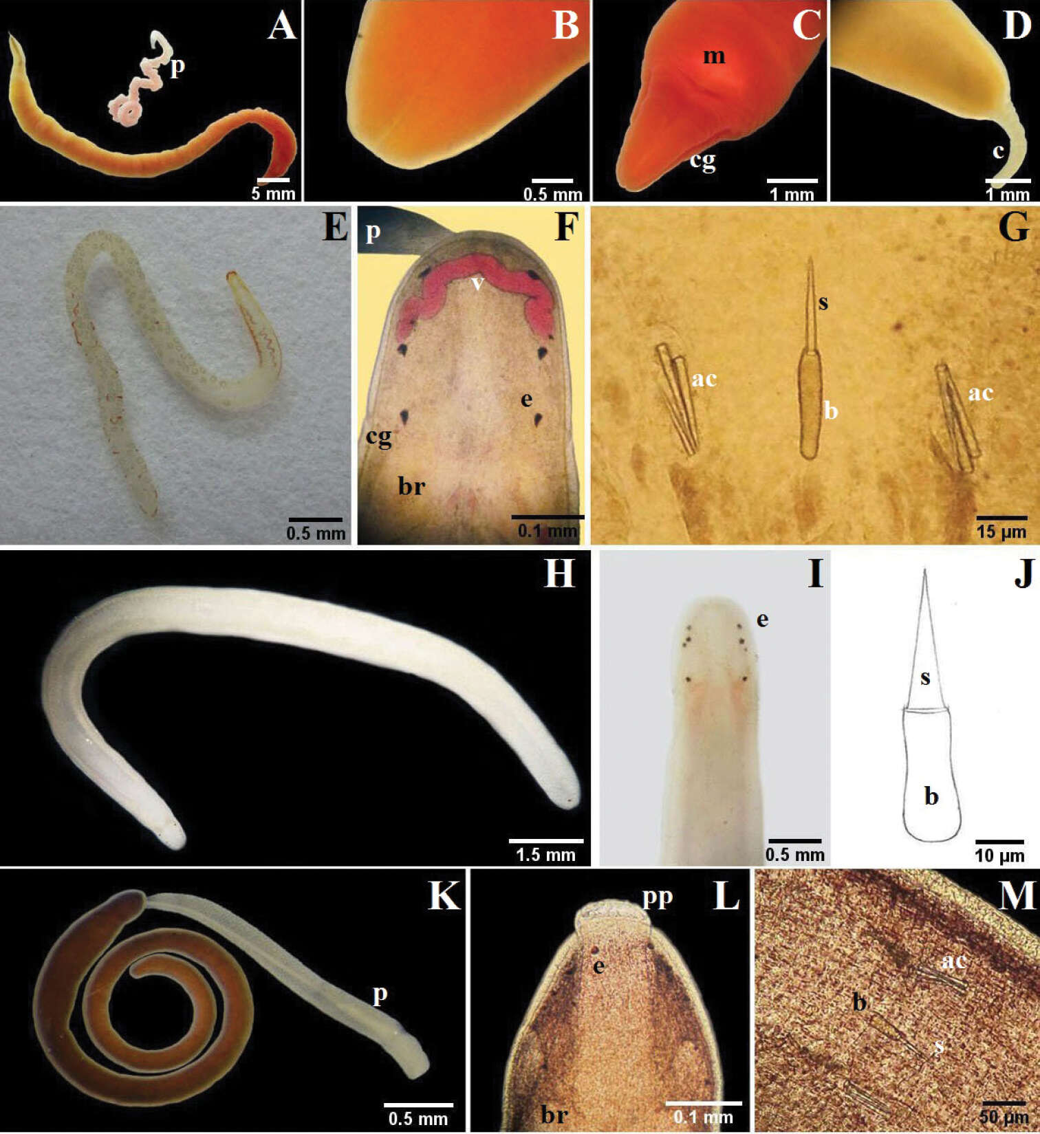
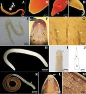
Figure 3.A–D Micrura ignea: A entire specimen, the worm has expulsed the proboscis B dorsal detail of the head C ventral detail of the head D detail of the tail E–G Amphiporus cruentatus: F dorsal detail of the head G detail of the stylets H–J Amphiporus cf. ochraceus: I dorsal detail of the head J drawing of the stylet K–M Amphiporus texanus: K entire worm L dorsal detail of the head M detail of the stylets. ac accessory stylet, b base of the stylet, br brain, c cirrus, cg cephalic grooves, e eyes, m mouth, p proboscis, pp proboscis pore, s sylet, v blood vessel.
-
Jaime Gonzalez-Cueto, Sigmer Quiroga, Jon Norenburg
Zookeys
Figure 3.A–D Micrura ignea: A entire specimen, the worm has expulsed the proboscis B dorsal detail of the head C ventral detail of the head D detail of the tail E–G Amphiporus cruentatus: F dorsal detail of the head G detail of the stylets H–J Amphiporus cf. ochraceus: I dorsal detail of the head J drawing of the stylet K–M Amphiporus texanus: K entire worm L dorsal detail of the head M detail of the stylets. ac accessory stylet, b base of the stylet, br brain, c cirrus, cg cephalic grooves, e eyes, m mouth, p proboscis, pp proboscis pore, s sylet, v blood vessel.
-
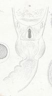
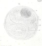
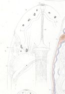
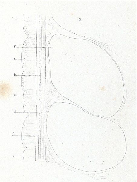
Plate13.19 Stylet-region of Amphiporus bioculatus
-
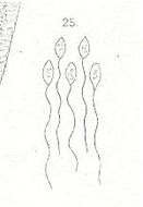
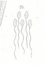
Plate7.25 Spermatozoa of Amphiporus bioculatus
-
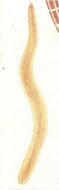
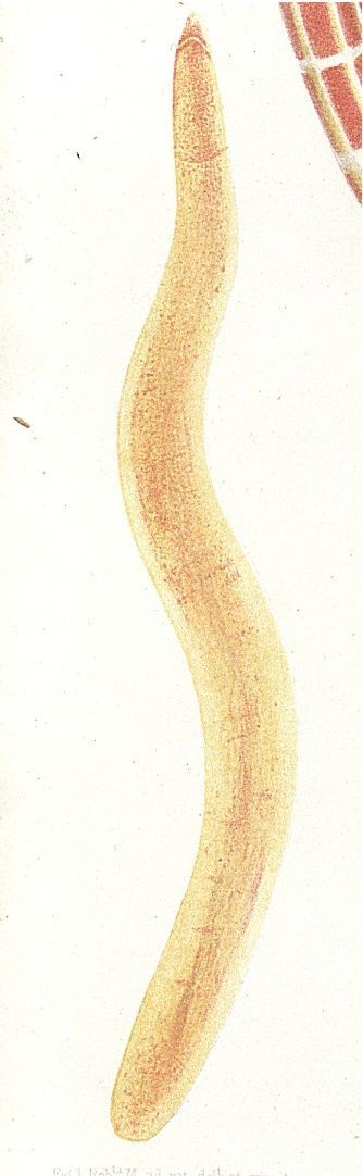
Plate8.3 Amphiporus bioculatus n. s.
-
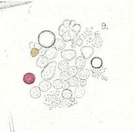
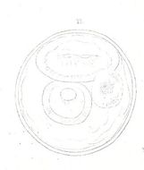
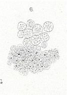
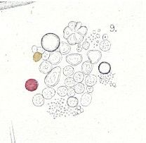
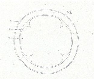
Plate10.9 Contents of the former, with oil-globules
-
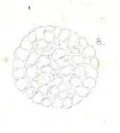
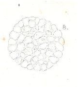
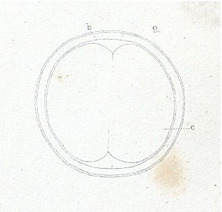
Plate10.8 One of the same slightly compressed glands
-
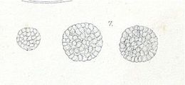
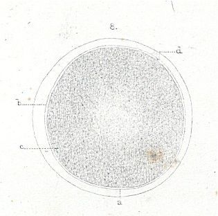
Plate10.7 Gland-cells from the wall of the digestive cavity of Amphiporus lactifloreus
-
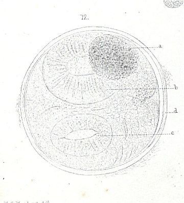
Plate7.14 Parasite extruded from the capsule
-
Plate7.13 The same ovum some hours afterwards, showing slight contraction of the discs
-
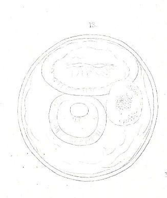
Plate7.12 Parasitic ovum immediately after removal
-
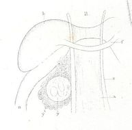
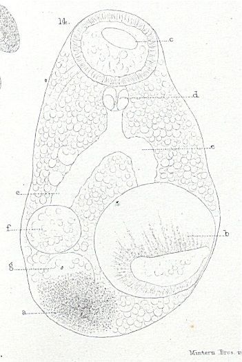
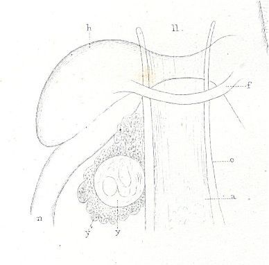
Plate7.11 Magnified view of a ganglionic region of a large one where a parasitic ovum lay imbedded in a granular lobulated mass
-
Plate7.2 Another specimen eight days older than the preceding
-
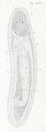
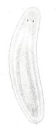
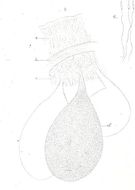
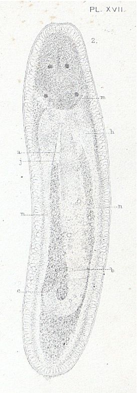
Plate7.1 A young specimen of Amphiporus lactifloreus on extrusion from the egg
-
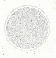
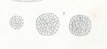
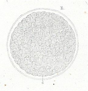
Plate6.12 Ovum just before the extrusion of the embryo
-
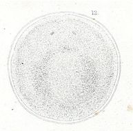
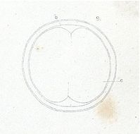
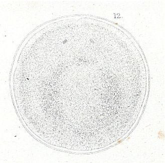
Plate6.11 Ovum of the same species in the mulberry-stage
-
Plate6.10 The same ovum a few hours later
-
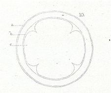
Plate6.9 The same ovum some hours after impregnation
-
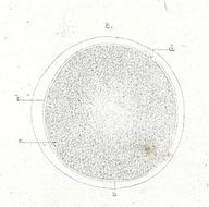
Plate6.8 Unimpregnated ovum of Amphiporus lactifloreus
-
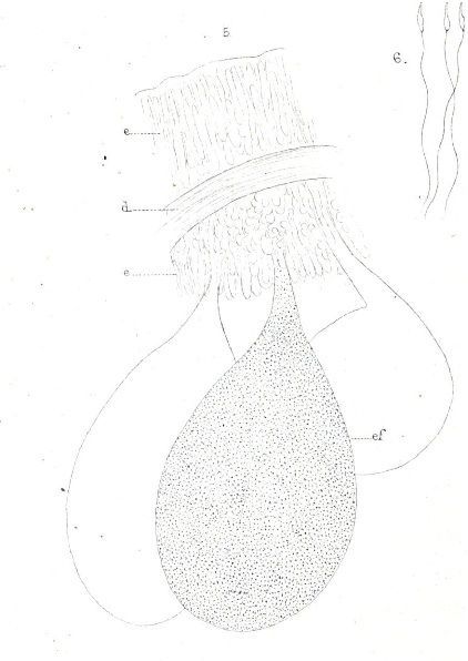
Plate6.5 Three sperm-sacs with a portion of the body-wall of Amphiporus lactifloreus
-
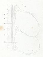
Plate6.2 Longitudinal section of the body-wall of Amphiporus lactfloreus, in a somewhat shriveled condition
-
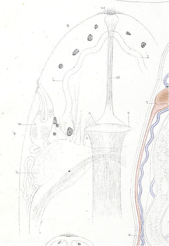
Plate5.6 Nerve-cells from a cephalic ganglion of Amphiporus lactifloreus
-
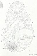
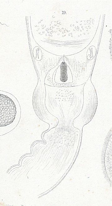
Plate5.4 Portion of the head of the same species considerably flattened