-
-
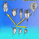
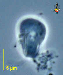
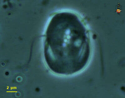
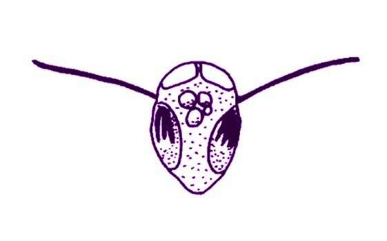
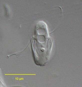
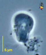
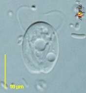
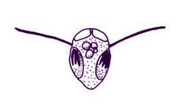
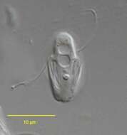
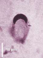
Octomitus is a diplomonad with a broadly pyriform cell body tapered posteriorly, (10-15 µm), bearing six anterior flagella deflected backwards and two posterior trailing flagella. The two anterior nuclei are bean-shaped, they face up and adjoin each other in their anterior part. A large endosome is present in the anterior lobe of the nuclei. The two sets of flagella emerge on each side of the anterior part of the body. The two recurrent flagella, accompanied by a sheath of reticulum, traverse the cell, forming a central axis before emerging as trailing flagella. Two spikes are located at the posterior between the two trailing flagella. There is no cytostomal opening at the emergence of the trailing flagella in contrast to Spironucleus. Anaerobic, parasitic or endocommensal in the intestine of vertebrates such as amphibians, caecum of rodents, rumen. Octomitus intestinalis from mice with two posterior flagella (phase contrast)
-
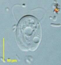
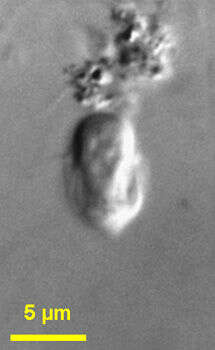
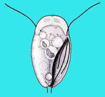
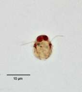
Trepomonas (tree-poe-moan-ass) is one of the free-living diplomonad flagellates. As with almost all diplomonads there are two anterior nuclei and two sets of flagella, one set associated with each nucleus. Different genera are distinguished largely by the numbers and relative lengths of the flagella. his genus has one long anterior flagellum in each group, and three very short ones lying within each of the lateral grooves. The anterior flagella are not evident here. Phase contrast
-
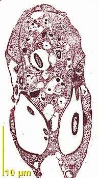
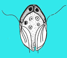
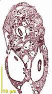
Octomitus is a diplomonad with a broadly pyriform cell body tapered posteriorly, (10-15 µm), bearing six anterior flagella deflected backwards and two posterior trailing flagella. The two anterior nuclei are bean-shaped, they face up and adjoin each other in their anterior part. A large endosome is present in the anterior lobe of the nuclei. The two sets of flagella emerge on each side of the anterior part of the body. The two recurrent flagella, accompanied by a sheath of reticulum, traverse the cell, forming a central axis before emerging as trailing flagella. Two spikes are located at the posterior between the two trailing flagella. There is no cytostomal opening at the emergence of the trailing flagella in contrast to Spironucleus. Anaerobic, parasitic or endocommensal in the intestine of vertebrates such as amphibians, caecum of rodents, rumen. Octomitus intestinalis from mice with two anterior nuclei, six antero-lateral flagella and two posterior flagella which traverse the cell axially (Giemsa staining)
-
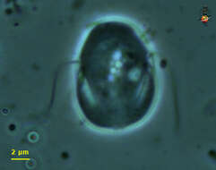
Trepomonas (tree-poe-moan-ass) is one of the free-living diplomonad flagellates. As with almost all diplomonads there are two anterior nuclei and two sets of flagella, one set associated with each nucleus. Different genera are distinguished largely by the numbers and relative lengths of the flagella. This genus has one long anterior flagellum in each group, and three very short ones lying within each of the lateral grooves. Differential interference contrast.
-
Trepomonas (tree-poe-moan-ass) is one of the free-living diplomonad flagellates. As with almost all diplomonads there are two anterior nuclei and two sets of flagella, one set associated with each nucleus. Different genera are distinguished largely by the numbers and relative lengths of the flagella. his genus has one long anterior flagellum in each group, and three very short ones lying within each of the lateral grooves. Phase contrast.
-
-
-
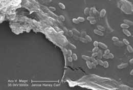
This scanning electron micrograph (SEM) of an untreated water specimen extracted from a wild stream mainly used to control flooding during inclement weather, revealed the presence of unidentified organisms, which included bacteria, protozoa, and algae. In this particular image, a protective biofilm had been inhabited by numbers of what appeared to be unidentified bacterial microorganisms.Created: 2009
-
With one long flagellum projecting from each groove on opposing sides of the cell. Encountered in organically very enriched sediments. Phase contrast optics.
-
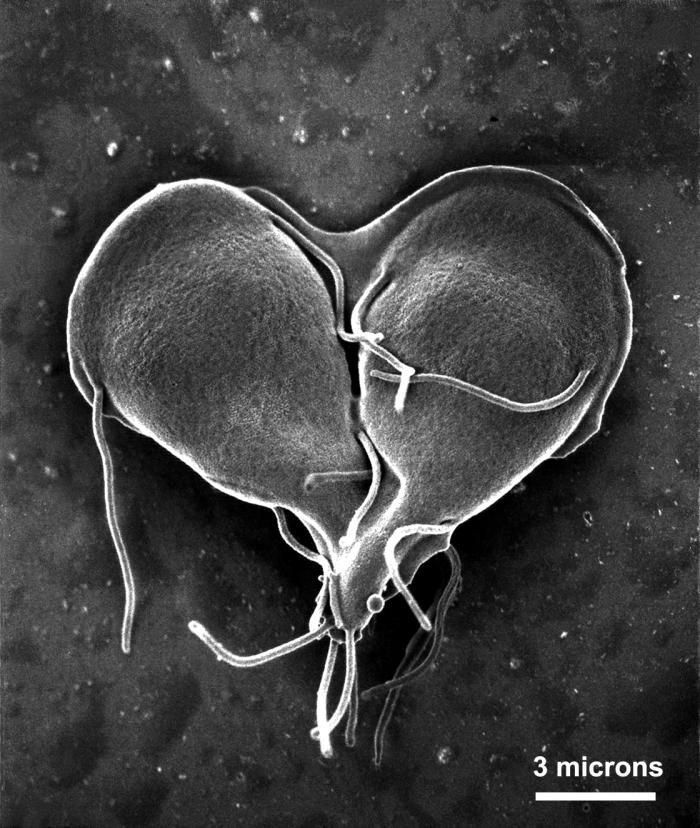
This scanning electron micrograph (SEM) depicted a Giardia lamblia protozoan that was about to become two, separate organisms, as it was caught in a late stage of cell division, producing a heart-shaped form.Created: 1999
-
With two long flagella projecting from the grooves on opposing sides of the cell. There are three other flagella in each groove, and one of these (left side) has also emerged from the groove. Phase contrast micrograph.
-
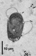
Trepomonas agilis: A diplomonad flagellate with a single prominant lateral flagella on each side. Three pairs of additional flagella lodged in the lateral grooves is also present, not visible in this image. This image was taken by Krishnakumar B. from one of the anaerobic bioreactors for organic rich wastewater treatment in Regional Research Laboratory-Trivandrum (CSIR-India).
-
Trepomonas (tree-poe-moan-ass) agilis Dujardin, 1841. Cell is ovoid, but S-shaped in cross section and is about 11 microns long. Two nuclei are located anteriorly. Two groups of flagella are inserted laterally at the end of each groove: two long flagella and six short flagella. The length of the long flagella was not measured, but the short falgella are less than half the cell length and lie in the grooves. The cell moves by swimming. Contractile vacuoles are seen. Rarely observed.
-
Trepomonas (tree-poe-moan-ass) agilis Dujardin, 1841. Cells are 8-12microns long, S-shaped in cross-section, ovoid, egg-shaped or elongate. Two opposed grooves run spirally, along the posterior two thirds to three quarters of the cell. Four flagella insert at the head of each groove. Of these, one flagellum, the same length as the cell or slightly shorter, is directed laterally. The other flagella are less than half the length of the cell, are directed posteriorly and lie within the groove. Two elongate nuclei are located anteriorly. The cytoplasm has a granular appearance, and exhibits cyclosis. Food vacuoles and empty vacuoles are scattered within the cell. Cells rotate smoothly as they swim.
-
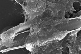
This scanning electron micrograph (SEM) of an untreated water specimen extracted from a wild stream mainly used to control flooding during inclement weather, revealed the presence of unidentified organisms, which included bacteria, protozoa, and algae. Visible in this particular image were a number of different microorganisms including elongated diatoms, and an amorphic gelatinous biofilm mass, which had enveloped amoeboid and bacterial organisms. For a colorized version of this image, see PHIL 11712.Created: 2009
-
Trepomonas agilis (Dujardin, 1841). Cell is ovoid, but S-shaped in cross section. Two nuclei are located anteriorly. Two groups of flagella are inserted laterally at the end of each groove: two long flagella and six short flagella. The short falgella are less than half the cell length and lie in the grooves (seen best here to viewer's left). Contractile vacuoles are seen.Collected from putifying sample from a freshwater pond near Boise, Idaho.DIC.
-
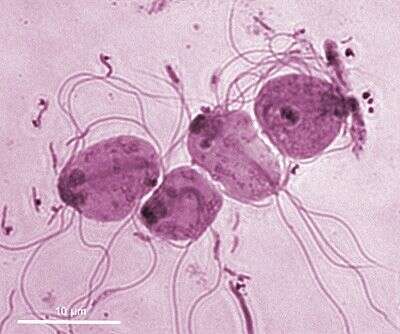
This is a scanning electron micrograph (SEM) of an in vitro Giardia lamblia culture, which had been cultivated in bile-free TYI-S-33 medium for 48 hrs, then incubated 24 hrs with 10 mg/ml bovine bile in order to stimulate cyst formation. This photograph contains both trophozoites, and a cluster of maturing cysts (bottom right). At far left, the two trophozoite-staged organisms are positionally situated opposite to one another, with the farthest left G. lamblia displaying its dorsal, or upper surface, and the protozoan to its immediate right, its ventral, or bottom surface.Created: 1999
-
Trepomonas agilis Dujardin, 1841. Cell is ovoid, but S-shaped in cross section. Two nuclei are located anteriorly (densley stained here). Two groups of flagella are inserted laterally at the end of each groove: two long flagella and six short flagella (seen here).Stained by the silver carbonate technique (see Foissner, W. Europ. J. Protistol., 27:313-330;1991). Collected from a putrifying sample from a freshwater pond near Boise, Idaho.Brightfield.
-
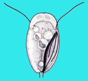
Diplomonad flagellate with a bilaterally compressed cell (5-30 µm) with two anterior nuclei, two lateral locomotory flagella, and two sets of three recurrent flagella situated in two posterior grooves. The anterior part of the cell is occupied by two crescent-shaped nuclei which abut on top. The two set of flagella are inserted on each side of the cell body near the equator at the base of each nucleus. The posterior half of the cell is grooved by two depressions or pockets each containing three recurrent flagella. The grooves are the site of ingestion of food, usually bacteria. One contractile vacuole forms in the middle part and discharges at the posterior end. Free-living in freshwater microaerophilic habitats.Image showing two lateral locomotory flagella (haematoxylin staining).
-
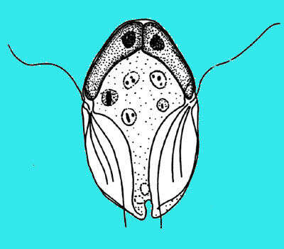
Diplomonad flagellate with a bilaterally compressed cell (5-30 µm) with two anterior nuclei, two lateral locomotory flagella, and two sets of three recurrent flagella situated in two posterior grooves. The anterior part of the cell is occupied by two crescent-shaped nuclei which abut on top. The two set of flagella are inserted on each side of the cell body near the equator at the base of each nucleus. The posterior half of the cell is grooved by two depressions or pockets each containing three recurrent flagella. The grooves are the site of ingestion of food, usually bacteria. One contractile vacuole forms in the middle part and discharges at the posterior end. Free-living in freshwater microaerophilic habitats. Image showing two two anterior crescent-shaped nuclei (haematoxylin)
-
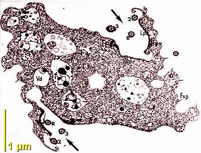
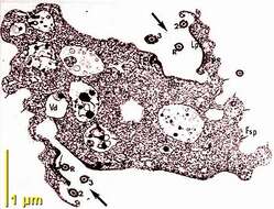
Transmission EM, longitudinal section showing the lateral insertion of the basal bodies/flagella and the two posterior cytostomal dimples.
-
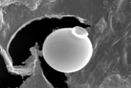
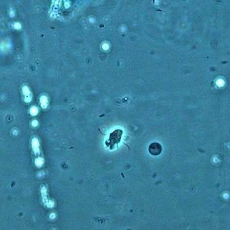
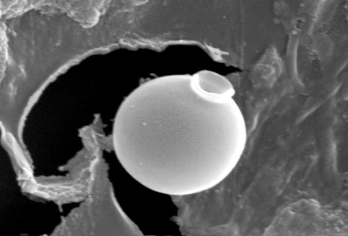
This scanning electron micrograph (SEM) of an untreated water specimen extracted from a wild stream mainly used to control flooding during inclement weather, revealed the presence of unidentified organisms, which included bacteria, protozoa, and algae. Clearly visible in the center of this image, was a exquisitely-formed unidentified round vescicle-shaped microorganism, which may have been algal, or diatomic. Shaped like an ancient Grecian urn, the almost perfectly rounded smooth, flawless surface was made even more beautiful given its delicate structure. For a colorized version of this image, see PHIL 11709.Created: 2009
-
Transmission EM, posterior transverse section showing the two opposite cytostomal openings containing three recurrent flagella.