-
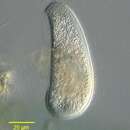
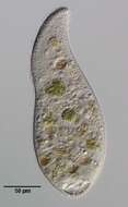
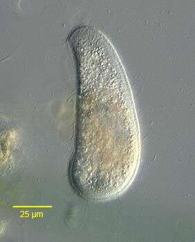
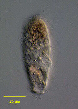
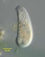
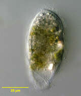
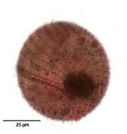
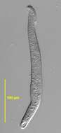
Bryophyllum vorax (Stokes, 1888) Kahl, 1931. Collected from a freshwater pond near Boise, Idaho. DIC
-
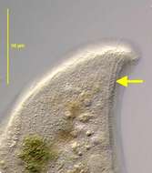
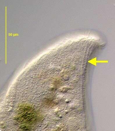
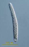
Spathidium (spa-thid-ee-um) moniliforme, the body is elongate, the posterior end is bluntly pointed or rounded but the anterior end is distinctively swollen - often fan-shaped and obliquely truncated. There is an ciliated apical ridge which is lined by toxicysts. The oral aperture is a slit that lies along the length of this ridge. The cilia are uniformly distributed in longitudinal parallel rows on both lateral surfaces. The macronucleus is highly variable, often elongate, ribbon-like or moniliform. The contractile vacuole is single and at the end of the cell. Spathidium feeds on other ciliates. It lives in fresh water ponds and lakes. This specimen was collected in a freshwater pond near Konstanz, Germany. This swimming cell is 250 microns long. Differential interference contrast.
-
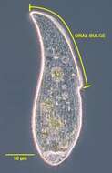
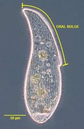
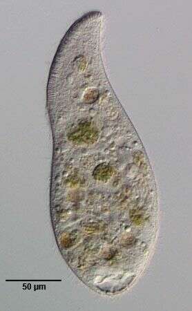
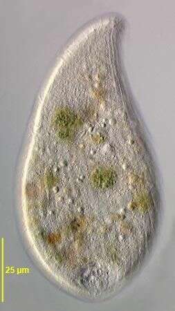
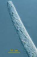
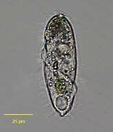
The long oral bulge (~50% of cell length) is one of the main distinguishing features of this subspecies of A. cultriforme. This specimen is somewhat stouter than the cells described by Foissner (Protistology 4 (1), 5-55 (2005) probably due to contraction after transfer from the culture dish to the slide. When observed undisturbed under the dissecting microscope the cells appear more slender.Phase contrast.
-
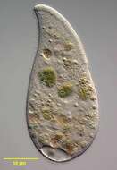
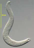
Portrait (left side) of the haptorid ciliate, Perispira ovum (Stein, 1859). The cell body is cylindrical to ovoid. The anterior end is slightly truncate. An unciliated cortical ridge makes a complete right-hand spiral the length of the body (seen here running obliquely across mid body). The slit-like cytostome, supported by fine trichites, is located at the anterior end of the cortical ridge. The uniform longitudinal somatic kineties spiral slightly. Densely packed food vacuoles and highly refractile cytoplasmic crystals often obscure the ellipsoid macronucleus. There is a single large terminal contractile vacuole posteriorly. Swims slowly. Collected from anoxic bottom sediments of slow flowing freshwater stream near Boise, Idaho march 2004. DIC optics
-
Spathidium (spa-thid-ee-um) moniliforme, the body is elongate, the posterior end is bluntly pointed or rounded but the anterior end is distinctively swollen - often fan-shaped and obliquely truncated. There is an ciliated apical ridge which is lined by toxicysts. The oral aperture is a slit that lies along the length of this ridge. The cilia are uniformly distributed in longitudinal parallel rows on both lateral surfaces. The macronucleus is highly variable, often elongate, ribbon-like or moniliform. The contractile vacuole is single and at the end of the cell. Spathidium feeds on other ciliates. It lives in fresh water ponds and lakes. This cell is squashed allowing ribbon-like macronucleus and the fan-like arrangement of toxicysts at the front of the cell to be seen. Differential interference contrast.
-
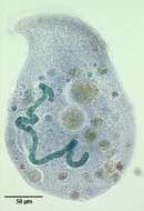
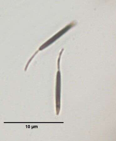
Six of the multiple (7-18) micronuclei are in the focal plane of this image. Stained by the methylgreen-pyroninY technique (see Foissner, W.Europ. J. Protistol.27:313-330;1991).Brightfield.
-
Portrait (right side) of the haptorid ciliate, Perispira ovum (Stein, 1859). The cell body is cylindrical to ovoid. The anterior end is slightly truncate. An unciliated cortical ridge makes a complete right-hand spiral the length of the body. The slit-like cytostome, supported by fine trichites, is located at the anterior end of the cortical ridge. The uniform longitudinal somatic kineties spiral slightly. Densely packed food vacuoles and highly refractile cytoplasmic crystals often obscure the ellipsoid macronucleus. There is a single large terminal contractile vacuole posteriorly. Swims slowly. Collected from anoxic bottom sediments of slow flowing freshwater stream near Boise, Idaho march 2004. DIC optics
-
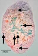
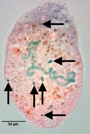
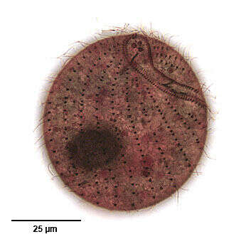
Band-form macronucleus of Arcuospathidum cultriforme scalpriforme (KAHL,1930) FOISSNER,2003.Stained by the methylgreen-pyroninY technique (see Foissner, W.Europ. J. Protistol.27:313-330;1991).Brightfield.
-
Portrait (right side) of the haptorid ciliate, Perispira ovum (Stein, 1859). The cell body is cylindrical to ovoid. The anterior end is slightly truncate. A narrow unciliated cortical ridge makes a complete right-hand spiral the length of the body. The slit-like cytostome, supported by fine trichites (seen well in this image), is located at the anterior end of the cortical ridge. The uniform longitudinal somatic kineties spiral slightly. Densely packed food vacuoles and highly refractile cytoplasmic crystals often obscure the ellipsoid macronucleus. There is a single large terminal contractile vacuole posteriorly. Swims slowly. Collected from anoxic bottom sediments of slow flowing freshwater stream near Boise, Idaho March 2004. DIC optics.
-
-
Portrait (lateral view) of the haptorid ciliate, Perispira ovum (Stein, 1859). The cell body is cylindrical to ovoid. The anterior end is slightly truncate. An unciliated cortical ridge makes a complete right-hand spiral the length of the body. The slit-like cytostome, supported by fine trichites, is located at the anterior end of the cortical ridge. The uniform longitudinal somatic kineties spiral slightly. Densely packed food vacuoles and highly refractile cytoplasmic crystals often obscure the ellipsoid macronucleus. There is a single large terminal contractile vacuole posteriorly. Swims slowly. Collected from anoxic bottom sediments of slow flowing freshwater stream near Boise, Idaho December 2004. Brightfield optics, closed condenser.
-
-
Portrait (left side) of the haptorid ciliate, Perispira ovum (Stein, 1859). The cell body is cylindrical to ovoid. The anterior end is slightly truncate. An unciliated cortical ridge makes a complete right-hand spiral the length of the body. The slit-like cytostome, supported by fine trichites, is located at the anterior end of the cortical ridge. The uniform longitudinal somatic kineties spiral slightly. Densely packed food vacuoles and highly refractile cytoplasmic crystals often obscure the ellipsoid macronucleus. There is a single large terminal contractile vacuole posteriorly. Swims slowly. Collected from anoxic bottom sediments of slow flowing freshwater stream near Boise, Idaho December 2004. DIC optics
-
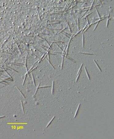
Stained by the silver carbonate technique (see Foissner, W.Europ. J. Protistol.27:313-330;1991).Brightfield.
-
Infraciliature (posterior apical view) of the haptorid ciliate, Perispira ovum (Stein, 1859). The cell body in vivo is cylindrical to ovoid. The longitudinal somatic kineties spiral slightly. An unciliated cortical ridge, bordered on either side by by a file of closely spaced kinetids, makes a complete right-hand spiral the length of the body. The posterior portion of this structure is seen here to the viewer's left. The three files of clavate cilia (dorsal brush) are seen at the viewr's upper right. The right-most kinety of the dorsal brush has longer cilia than the two kineties to it's left. Collected from anoxic bottom sediments of slow flowing freshwater stream near Boise, Idaho December 2004. Stained by the silver carbonate technic (see Foissner, W. Europ. J. Protistol., 27:313-330;1991).Brightfield.
-
-
Infraciliature (ventrolateral view) of the haptorid ciliate, Perispira ovum (Stein, 1859). The cell body in vivo is cylindrical to ovoid. The longitudinal somatic kineties spiral slightly. An unciliated cortical ridge, bordered on either side by by a file of closely spaced kinetids, makes a complete right-hand spiral the length of the body. The anterior portion of this structure is seen here. The cytostome is located at the anterior end of the spiral ridge. The cytostome is supported by trichites (not seen here).Collected from anoxic bottom sediments of slow flowing freshwater stream near Boise, Idaho December 2004. Stained by the silver carbonate technic (see Foissner, W. Europ. J. Protistol., 27:313-330;1991).Brightfield.
-
2 of the three dorsal brush rows are seen in this image.DIC.
-
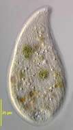
Portrait of the haptorid ciliate, Perispira ovum (Stein, 1859). The cell body is cylindrical to ovoid. The anterior end is slightly truncate. An unciliated cortical ridge (not well seen here) makes a complete right-hand spiral the length of the body. The slit-like cytostome, supported by fine trichites, is located at the anterior end of the cortical ridge. The uniform longitudinal somatic kineties spiral slightly. There are thre files of clavate (club-shaped) cilia forming a dorsal brush. The right-most of these (visible here to the viewer's upper right) hhas longer cilia than the two kineties to it's right. The cytoplasm in this individual is densely packed chloroplasts from ingested euglenae. These are probably "kleptoplasts". Kleptoplasts are plastids from ingested prey that are maintained in the cytoplasm and not digested. Perispira ovum may also sequester mitochondria from its prey in similar fashion (Johnson, PW et al. J. Euk. Microbiol.42:323-335,1995). There is a single large terminal contractile vacuole posteriorly. Collected from anoxic bottom sediments of slow flowing freshwater stream near Boise, Idaho march 2004. DIC.
-
-
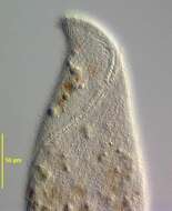
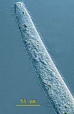
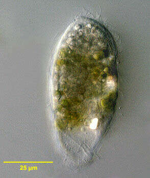
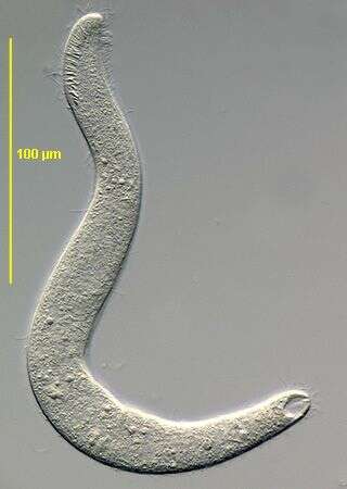
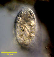
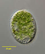
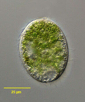
Collected from a non-flooded Petri dish culture of topsoil from a public park in Boise, Idaho. November 2006.DIC.
-
-
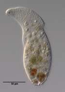
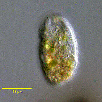
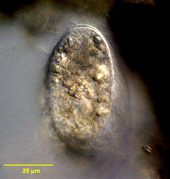
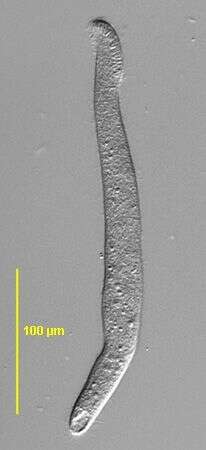
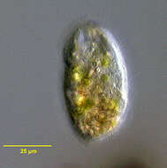
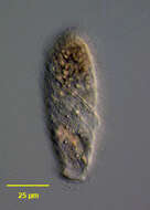
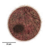
Portrait of Arcuospathidium (FOISSNER,1984). Collected from a non-flooded Petri dish culture of topsoil from a public park in Boise, Idaho. November 2006.DIC.
-