-
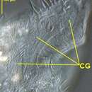
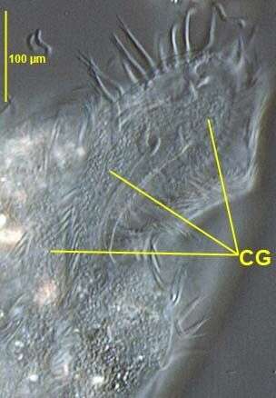
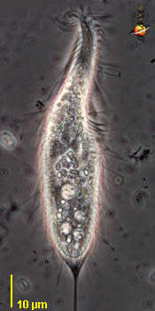
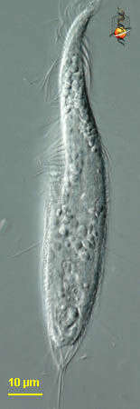
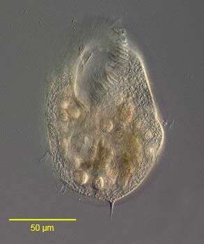
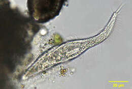
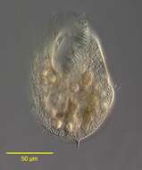
in vivo view of the amphisiellid hypotrich Pseudouroleptus caudatus (HEMBERGER,1985) showing colorless cortical granules (CG).Specimen from rewetted soil sample from grass lawn of a public park in Boise,Idaho.January 2007.DIC.
-
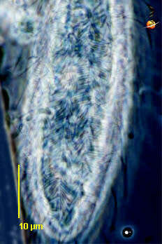
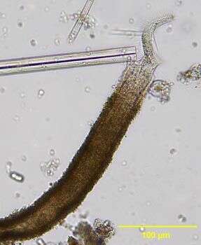
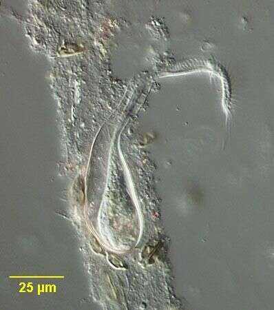
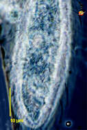
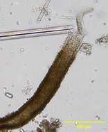
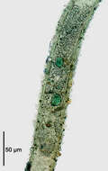
Stichotricha (stike-o-trike-a), un-named species. The body of the unidentified hypotrich ciliate has a broader posterior portion, and a narrowed anterior region. This is a detail of the cell surface showing the numerous long ectosymbiotic bacteria. Phase contrast.
-
-
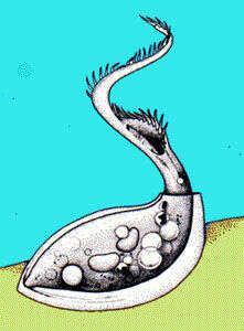
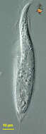
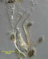
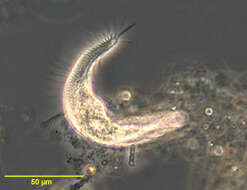
Portrait of the stichotrichine ciliate, Chaetospira remex (Hudson,1875; Kahl,1932). This species occupies a long, sometimes branched tubular lorica into which it intermittently retracts (as seen in this image). The lorica is attached to the substratum. C. muelleri has a flask-shaped lorica. The cell body is slender,elongate and very contractile. The corkscrew shaped anterior bears a prominent adoral zone of membranelles along the peristome. The somatic ciliature is reduced to right and left marginal and two ventral files of short cirri which spiral down the body. The macronucleus is bipartite. the contractile vacuole is in mid-body between the two macronuclei. Feeds mainly on bacteria, flagellates and diatoms. Collected from a freshwater pond near Boise, Idah May 2004. DIC optics.
-
Vallibona, Valencia, Spain
-
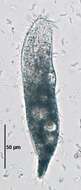
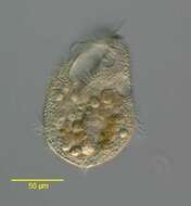
in vivo view of the amphisiellid hypotrich Pseudouroleptus caudatus (HEMBERGER,1985) .Specimen from rewetted soil sample from grass lawn of a public park in Boise,Idaho.January 2007.Brightfield.
-
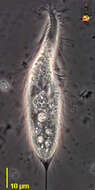
Stichotricha (stike-o-trike-a), un-named species. The body of the unidentified hypotrich ciliate has a broader posterior portion, and a narrowed anterior region. The adoral zone of membranelles makes about one complete tour of the anterior part, winding down from the anterior to the cytostome located in the anterior part of the broader part of the cell. There are locomotor cirri over the body (not easily seen here), and also stiff projecting cilia / cirri. Some structures form a bracing structure to support the two very long cilia which extend 50 or 60 microns Phase contrast micrograph.
-
Portrait of Chaetospira, a loricate hypotrich ciliate. Elongate anterior end with prominent adoral zone of membranelles has a corkscrew configuration, distinguishing this genus from the similar Stichotricha. Usually only anterior portion protrudes and organism quickly retracts completely into lorica when disturbed. From freshwater pond near Boise, Idaho. Brightfield.
-
Portrait of the stichotrichine ciliate, Chaetospira remex (Hudson,1875; Kahl,1932). Slightly squashed. This species occupies a long, sometimes branched tubular lorica into which it intermittently retracts. The lorica is attached to the substratum. C. muelleri has a flask-shaped lorica. The cell body is slender,elongate and very contractile. The corkscrew shaped anterior bears a prominent adoral zone of membranelles along the peristome (seen well here). The somatic ciliature is reduced to right and left marginal and two ventral files of short cirri which spiral down the body. The left marginal and one of the ventral cirral files are seen here.The macronucleus is bipartite (not visible in this image). the contractile vacuole is in mid-body between the two macronuclei. Feeds mainly on bacteria, flagellates and diatoms. Collected from a freshwater pond near Boise, Idah May 2004. DIC optics.
-
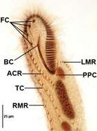
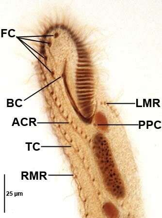
Pseudouroleptus caudatus Hemberger,1985. FC=frontal cirri.BC=buccal cirrus. PPC=postperistomial cirrus. RMR,LMR=right and left marginal cirral rows.ACR=amphisiellid median cirral row. TC=transverse cirral row.Specimen from rewetted soil sample from grass lawn of a public park in Boise,Idaho.January 2007.Protargol impregnation,Wilbert modification (see Foissner, W. Europ. J. Protistol., 27:313-330;1991).Brightfield.
-
Stichotricha (stike-o-trike-a), un-named species. The body of the unidentified hypotrich ciliate has a broader posterior portion, and a narrowed anterior region. The adoral zone of membranelles can be seen to the right of the anterior-most part of the cell and then to the left near the cytostome located in the anterior part of the broader part of the cell - showing that it makes about one complete turn around the body. There is a sheath of material (ectosymbiotic bacteria?) which supports the caudal cilia. Differential interference contrast.
-
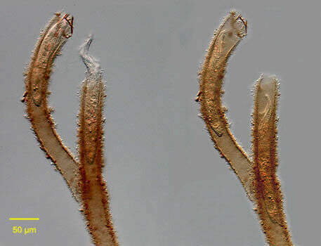
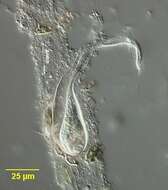
Chaetospira muelleri (LACHMAN,1856). The corkscrew shaped highly contractile anterior end of the cell is seen protriding through the opening of the flask-shaped lorica. Collected from tidal pools at Alki Beach, Seattle, Washington 47°35â41.25âN 122°23â19.60âW.January,2006. DIC.
-
The stichotrichine ciliate, Chaetospira remex (Hudson,1875; Kahl,1932) stained with methyl green-pyronin to demonstrate the bipartite macronucleus. The animal is completely withdrawn into the tubular lorica. The two round macronuclei are stained green. The micronucleus is not seen in this image. The illustration in Kahl's compendium incorrectly depicts C. remex with a single macronucleus (Kahl,A.; Die Tierwelt Deutschlands und der angrenzenden Meeresteile. Teil 25 [Urtiere oder Protozoa I: Wimpertiere oder Ciliata (Infusoria) 3. Spirotricha. Germany:Verlag von Gustav Fischer. 1932, p. 542).Collected from a freshwater pond near Boise Idaho May 2004. Brightfield illumination with closed condenser.
-
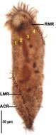
Dorsal infraciliature of Pseudouroleptus caudatus (HEMBERGER,1985) .RMR,LMR right and left marginal cirral rows.ACR=amphisiellid median cirral row. 1-4+dorsal kineties.Specimen from rewetted soil sample from grass lawn of a public park in Boise,Idaho.January 2007.Protargol impregnation,Wilbert modification (see Foissner, W. Europ. J. Protistol., 27:313-330;1991).Brightfield.
-
Stichotricha (stike-o-trike-a), un-named species. The body of the unidentified hypotrich ciliate has a broader posterior portion, and a narrowed anterior region. The adoral zone of membranelles makes about one complete tour of the anterior part, winding down from the anterior to the cytostome located in the anterior part of the broader part of the cell. This image illustrates the two very long cilia which extend 50 or 60 microns Phase contrast micrograph.
-
Chaetospira muelleri (LACHMAN,1856). This individual is completely retracted into the the flask-shaped lorica. Collected from tidal pools at Alki Beach, Seattle, Washington 47°35â41.25âN 122°23â19.60âW.January,2006. DIC.
-
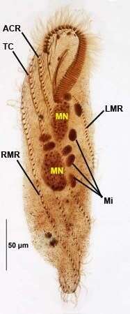
Ventral infraciliature of Pseudouroleptus caudatus (HEMBERGER,1985) .RMR,LMR right and left marginal cirral rows.TC=transverse cirral row.ACR=amphisiellid median cirral row.MN=two macronuclear nodules.Mi=multiple (up to eight) dense micronuclei.Specimen from rewetted soil sample from grass lawn of a public park in Boise,Idaho.January 2007.Protargol impregnation,Wilbert modification (see Foissner, W. Europ. J. Protistol., 27:313-330;1991).Brightfield.
-
-
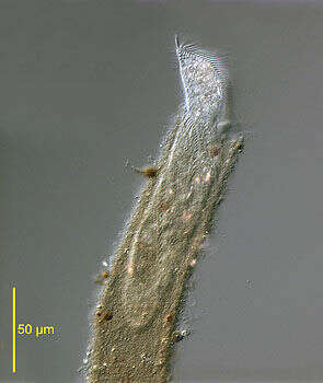
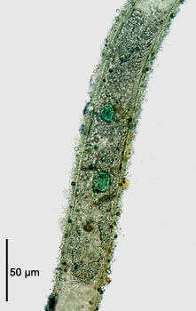
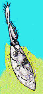
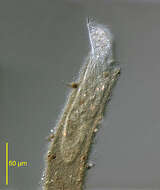
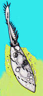
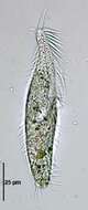
Stichotricha, hypotrich ciliate usually located in a flocculent mucoid mass adhering to submerged vegetation or other debris. The adoral zone of membranelles (AZM) is supported on a narrow and twisted anterior projection. Rows of cirri follow a curving path on the body.
-
Stichotricha, hypotrich ciliate usually located in a flocculent mucoid mass adhering to submerged vegetation or other debris. The adoral zone of membranelles (AZM) is supported on a narrow and twisted anterior projection. Rows of cirri follow a curving path on the body. Phase contrast optics.
-
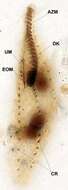
Stichotricha aculeata (WRZEÅNIOWSKI,1866).AZM=adoral zone of membranelles;DK=dorsal kinety;CR=2 of the 4 cirral rows;UM=undulating membrane;EOM=endoral membrane.Protargol protocol A (see Foissner, W. Europ. J. Protistol., 27:313-330;1991).Brightfield.
-
Stichotricha aculeata (WRZEÅNIOWSKI,1866).Brightfield,closed condenser.
-
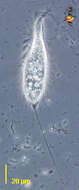
Portrait of the stichotrichine ciliate Hypotrichidium conicum (Ilowaisky, 1921). Collected from a freshwater pond near Boise, Idaho. July 2005. DIC.
-
in vivo portrait of the stichotrichine ciliate, Hypotrichidium conicum (Ilowaisky, 1921). Collected from a freshwater pond near Boise, Idaho. july 2005. DIC.