-
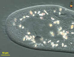
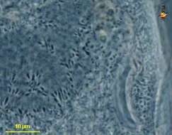
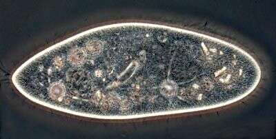
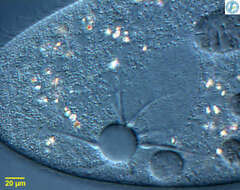
Phase contrast micrograph showing clearly one contractile vacuole with five filled channels.
-
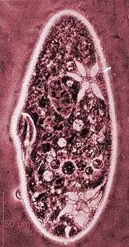
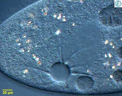
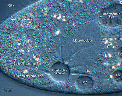
Paramecium with its iconic contractile vacuole. The central vacuole is surrounded by an array of collecting canals which collect fluid from the cytoplasm and feed it into the vacuole. The region of the canals near the vacuole are distensible ampullae. From Lake Donghu, China. Differential interference contrast micrograph.
-
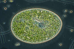
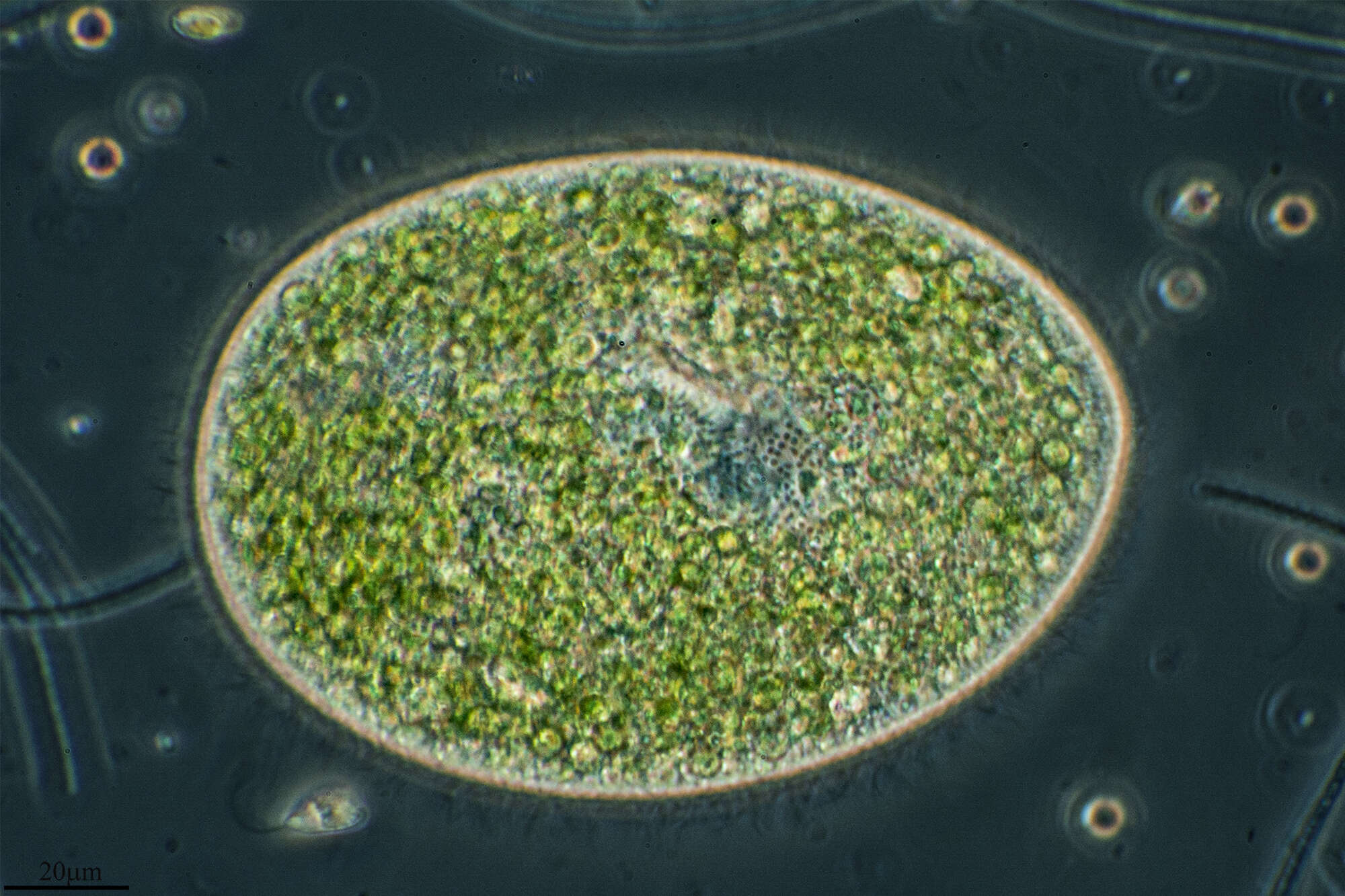
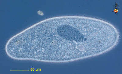
Paramecium (aurelia) (par-a-mee-see-um) is a very familiar genus of ciliates. They eat bacteria and have the mouth recessed in a buccal cavity, and the cell is often shaped with a scoop leading to the mouth. There are cilia all over the body with a caudal tuft of longer cilia at the back of the body. Usually with a layer of extrusomes (trichocysts) under the cell surface and a large oval macronucleus. Contractile vacuoles star-shaped. This species is P. aurelia, one of the smaller spindle-shaped (morpho)species. The (morpho) species is best distinguished by the presence of two small micronuclei pressed up against the macronucleus. Phase contrast.
-
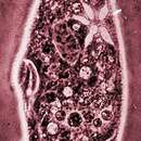
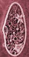
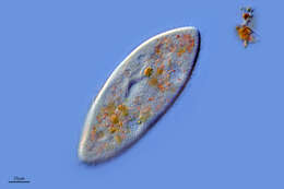
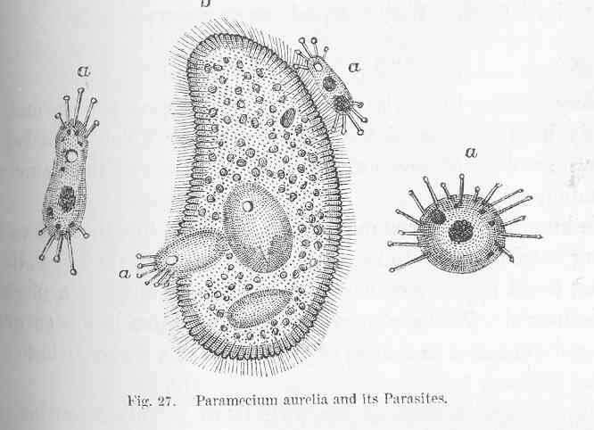
Paramecium aurelia and its Parasites.
-
Campillos, Andalusia, Spain
-
Herrera de Soria, Castille and Leon, Spain
-
Cabanas De Sayago, Castille and Leon, Spain
-
Caada del Hoyo, Castilla-La Mancha, Espaa
-
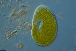
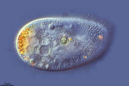
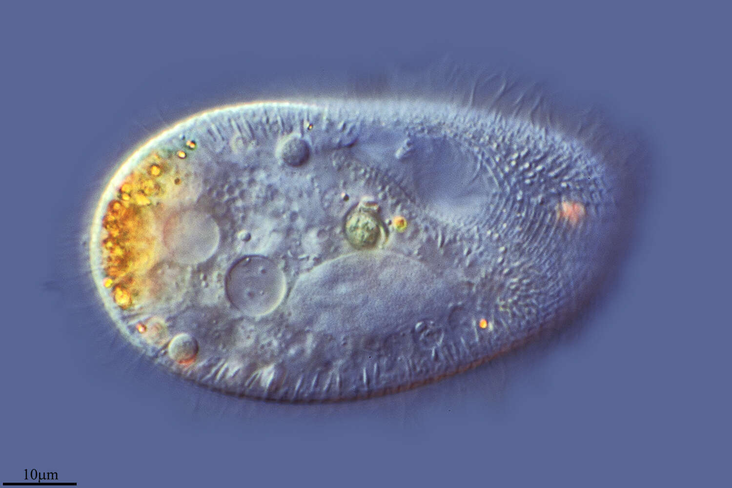
The Paramecium, shown in this image, was flattened slightly to make the cell components more visible. The cell is covered with hundreds of cilia. The dark structure upper right is the macronucleus.
-
Paramecium (aurelia) (par-a-mee-see-um) is a very familiar genus of ciliates and this (morpho) species is best distinguished by the presence of two small micronuclei pressed up against the macronucleus. They can be seen here to the north of the nucleus. Differential interference contrast.
-
Villoslada de Cameros, La Rioja, Spain
-
Ribadelago, Castille and Leon, Spain
-
Caada del Hoyo, Castilla-La Mancha, Espaa
-
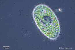
Paramecium with its iconic contractile vacuole. The central vacuole is surrounded by an array of collecting canals which collect fluid from the cytoplasm and feed it into the vacuole. The region of the canals near the vacuole are distensible ampullae. This image has been annotated by Marc Perkins at Orange Coast College (a community college in Costa Mesa, California).
-
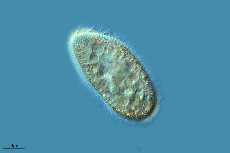
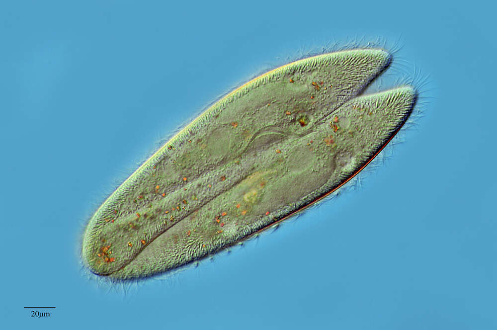
Paramecium (aurelia) (par-a-mee-see-um) is a very familiar genus of ciliates and this (morpho) species is best distinguished by the presence of two small micronuclei pressed up against the macronucleus. This image shows the peniculi or compound ciliary organelles in the mouth. Phase contrast.
-
-
Herrera de Soria, Castille and Leon, Spain
-
-
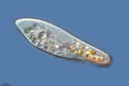
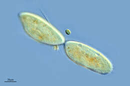
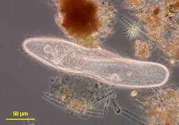
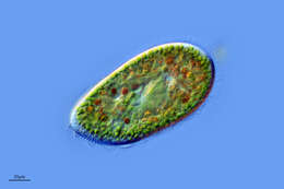
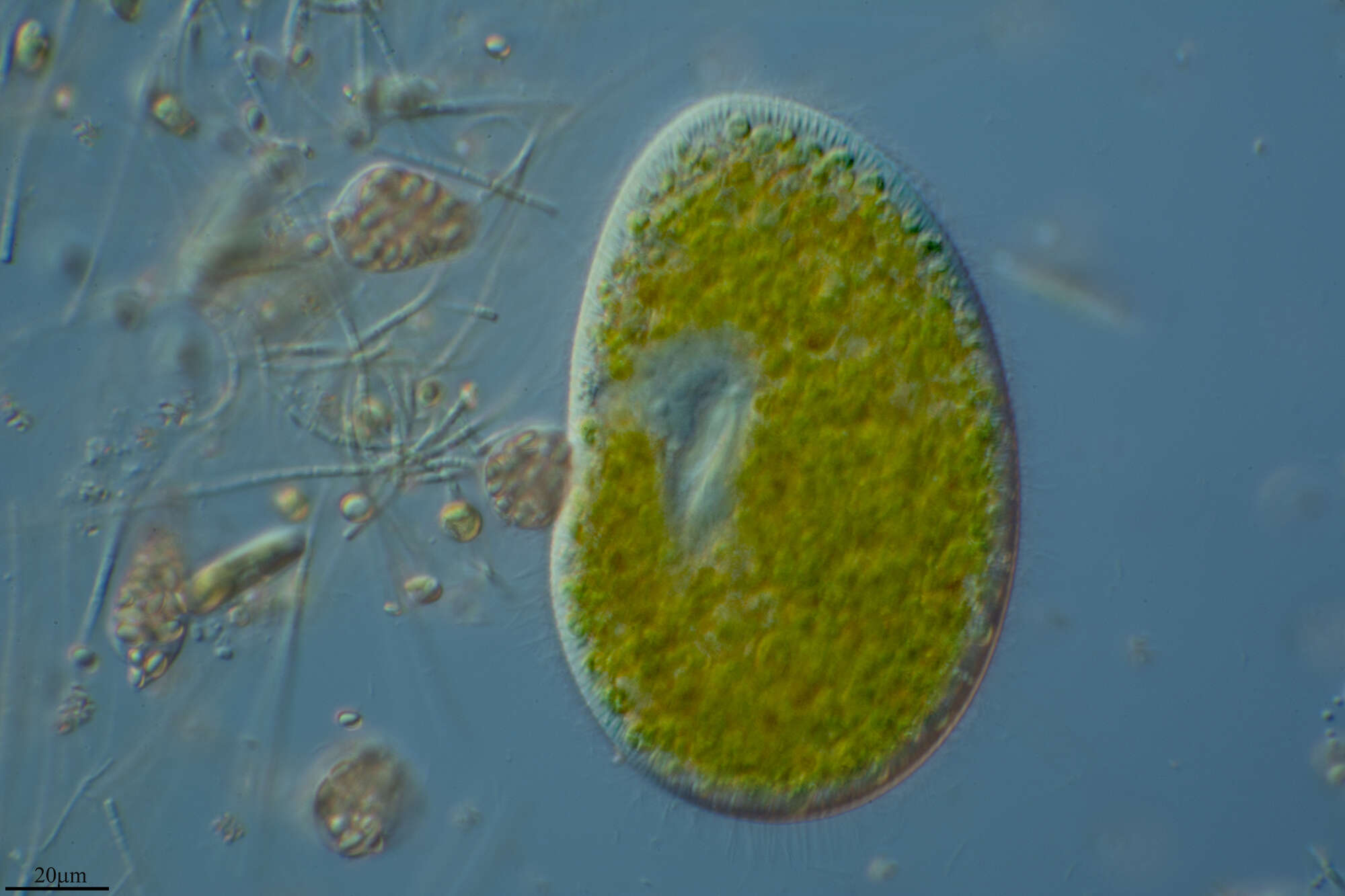
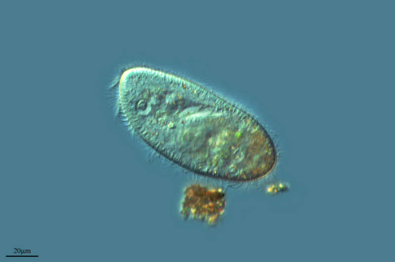
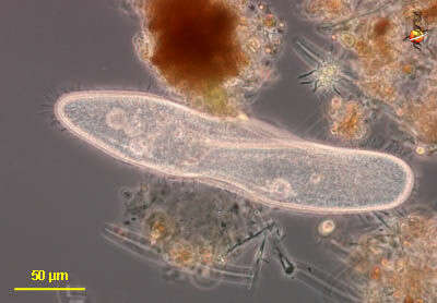
Paramecium (caudatum) (par-a-mee-see-um) is a very familiar genus of ciliates. They eat bacteria and have the mouth recessed in a buccal cavity, and the cell is often shaped with a scoop leading to the mouth. There are cilia all over the body with a caudal tuft of longer cilia at the back of the body. Usually with a layer of extrusomes (trichocysts) under the cell surface and a large oval macronucleus. Contractile vacuoles star-shaped. This species is P. caudatum, and was photographed with the cell pushing itself into some debris. This is the normal feeding behaviour of this genus. Phase contrast.
-
Paramecium (aurelia) (par-a-mee-see-um) is a very familiar genus of ciliates and this (morpho) species is best distinguished by the presence of two small micronuclei pressed up against the macronucleus. They can be seen here to the north of the nucleus. Phase contrast.
-
Castille and Leon, Spain
-
Ribadelago de Franco, Castilla y Len, Espaa
-
Cabanas De Sayago, Castille and Leon, Spain
-
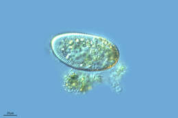
Paramecium (caudatum) (par-a-mee-see-um) is a very familiar genus of ciliates. They eat bacteria and have the mouth recessed in a buccal cavity, and the cell is often shaped with a scoop leading to the mouth. There are cilia all over the body with a caudal tuft of longer cilia at the back of the body. Usually with a layer of extrusomes (trichocysts) under the cell surface and a large oval macronucleus. Contractile vacuoles star-shaped. Most species are elongate, although this particular individual has been squashed so that we can see the nuclei. The (morpho)species are distinguished by morphology of the nuclei. This species is P. caudatum, which a single rather globular macronucleus lying alongside (4 o clock) the macronucleus. Phase contrast.