Comprehensive Description
provided by Smithsonian Contributions to Zoology
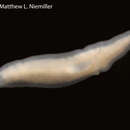
Sphalloplana (Speophila) hubrichti (Hyman, 1945)
Speophila hubrichti Hyman, 1945:479.
Sphalloplana hubrichti.—Mitchell, 1968:615.
Sphalloplana (Speophila) pricei.—Carpenter, 1971:1284 [in part].
TYPE MATERIAL.—Holotype, whole mount, AMNH 320. Paratypes, whole mount and sets of serial sections, AMNH 321–324 and 703 from Kolms Cave; whole mount and set of serial sections, AMNH 704, from Morrisons Cave.
EXTERNAL FEATURES (Figures 9, 25).—This is a rather large species, measuring up to 20 mm in length and 3 mm in width, in life of purely white color. The head is truncate, with slightly bulging frontal margin, frequently showing a median notch at the site of the adhesive organ. The lateral edges of the head are rounded, without auricular projections, but somewhat protruding laterally. The adhesive organ is visible in the living specimen as an opaque spot. The pharynx is situated behind the middle of the body and its length is about one-eighth the body length. The copulatory organs occupy the anterior half of the postpharyngeal region. The testes are visible in life as transparent spots, arranged in a pair of zones, several testes wide, to the right and left of the midline, each zone occupying the posterior half of the prepharyngeal region, extending posteriorly to the level of the anterior part of the pharynx. The intestinal zone ends anteriorly in a transversal border, not showing any V-shaped formation.
ANATOMY.—Apart from the materials of this species deposited in the American Museum of Natural History and in the United States National Museum, I had an opportunity of studying several live specimens from the type-locality in Missouri and from localities in Illinois.
The adhesive organ is a deep pit with irregularly folded walls and is provided with a complex musculature, as described by Hyman (1945:479).
The reproductive system was examined in the paratype slides and in 12 sets of serial sections preserved by a better method than that used by L. Hubricht for his original collection. The testes are of moderate size, dorsal or subdorsal. The ovaries are located usually at the level of the third or fourth lateral branch of the anterior intestinal ramus. Hyman's description of the copulatory complex needs some emendations. The genital pore (Figure 58, gp) leads into a rather small common genital atrium that connects anteriorly with the male atrium (am) and dorsally with the widened outlet (v) of the copulatory bursa. The penis consists of a rounded, muscular bulb and a large, generally finger-shaped papilla. Each vas deferens (vd), after expanding to form the convoluted spermiductal vesicle, enters the penial bulb ventrolaterally, proceeds toward the midline and opens into a rather large seminal vesicle (vs). What Hyman (1945:480) describes as “narrow ejaculatory duct” is actually the anteroposteriorly compressed, rather voluminous seminal vesicle that extends laterally to both sides. The epithelial lining of this vesicle is of a glandular nature, with club-shaped cells protruding into the lumen. In the median section, the vesicle generally arches from the anteroventral part of the bulb dorsally and posteriorly and, at the transition between bulb and papilla, connects with a duct that runs through the axis of the papilla to open at its tip. This canal, the true ejaculatory duct (de), is lined with a normal, nonglandular epithelium and varies considerably in its width and shape. It appears to have no surrounding muscular layer. The outer surface of the penis papilla is covered by a cuboidal epithelium, below which there is a well-developed layer of fine circular fibers, followed by a layer of coarser longitudinal muscles. In some specimens, the tip of the penis papilla was invaginated into the ejaculatory duct (Figure 59), so that the distal part of the duct showed the same muscular layers as the outer covering of the papilla. It is difficult to decide whether this invagination corresponds to the shape of the resting male organ that may extend at the moment of preservation by a contraction of the musculature of the penis papilla.
The spaceous male atrium (am) duplicates the shape of the penis. At its posterior end it narrows and connects with the small common atrium. The copulatory bursa (b) shows no peculiarities. Its outlet, the bursal duct or stalk (bd), begins as a narrow, straight canal running posteriorly above the penis bulb, then gradually widens, becomes more convoluted, and finally bends downward as an expanded section (v) situated to the left of the midline.
The two oviducts or ovovitelline ducts, which accompany the ventral nerve cord, ascend dorsally and medially at the level of the copulatory complex, each giving off a short posterior vitelline duct running toward the yolk glands of the tail region. The oviducts unite in the space above the male atrium and proceed posteroventrally as common oviduct (odc), provided with the usual eosinophilic shell glands, which opens into the common atrium from the dorsal side.
None of the epithelia of the copulatory complex are infranucleate.
DISTRIBUTION AND ECOLOGY.—Sphalloplana hubrichti inhabits a number of caves and springs in Missouri and Illinois. The following localities may be taken as habitats of the species.
MISSOURI. JEFFERSON COUNTY: Spring near Kimmswick and spring near Selma, collected by Leslie Hubricht in June 1937 (Hyman, 1945:480). SAINTE GENEVIEVE COUNTY: Kolms Cave (this is the correct spelling, rather than Kohn's Cave as Hyman indicates), type-locality of the species; many specimens collected by Leslie Hubricht in June 1937 (Hyman, 1945:480); about 35 specimens collected by Leslie Hubricht 13 September 1941 (alcohol material, USNM 50907); 6 specimens collected by Jerry Lewis and Margaret Meister 21 July 1974 and 12 April 1975.
ILLINOIS. JACKSON COUNTY: A walled spring in Happy Hollow, Fountain Bluff, south of Gorham, a total of 17 specimens collected by Jerry Lewis and Margaret Meister 10 February, 2 March, 14 April, 14 September, and 20 October 1974. MONROE COUNTY: Burksville Cave (also called Morrisons Cave, Eckerts Cave, Illinois Caverns, or Mammoth Cave of Illinois), collected by Leslie Hubricht, June 1937 (Hyman, 1945:480); about 25 specimens collected by Jerry Lewis 24 November 1974, occurring together with 2 species of Asellus and 4 species of amphipods. UNION COUNTY: Richs Cave, east of Cobden, 5 specimens, collected by Jerry Lewis, 2 February 1974, besides Phagocata gracilis (Haldeman).
Other distributional data for S. hubrichti given in the literature either need reexamination or are misidentifications caused by Hyman's defective description of the species. Beatty's (1966:10) “Cave near Cobden” is probably Richs Cave in Union County, Illinois, mentioned above. Beatty's Tom Moore Cave, Perry County, Missouri has Macrocotyla lewisi rather than S. hubrichti (see Kenk, 1975:333). McRitchie's (1959:20, 21) localities in Tennessee are probably all erroneous: the species occurring in the spring at Stokes Lane in Nashville is S. chandleri, described in this paper (p. 21). Specimens from the spring “at junction of U.S. routes 70N and 70S, Cheatham County,” and from Herring Cave, near Lascassas, Rutherford County, are not further described by McRitchie; S. chandleri from the Stokes Lane spring in Nashville is also mentioned by Darlington and Chandler (1972:160) and by Chandler (1972:62) as “Speophila hubrichti.”
TAXONOMIC POSITION.—By the development of a conspicuous adhesive organ, S. hubrichti belongs to the subgenus Speophila. Carpenter (1970:85) placed the species in synonymy with S. pricei, from which it may be distinguished in life by a slightly different shape of the head and by the transverse or rounded anterior border of the intestinal zone (which in S. pricei shows a V-shaped formation) and in the anatomy by the location of the testes (dorsal in S. hubrichti, dorsal and ventral in S. pricei). From the very similar S. chandleri it differs by the shape of the anterior intestinal border and by the entry of the vasa deferentia into the penis bulb (ventrolateral in S. hubrichti, dorsal after recurving in S. chandleri). The remaining American species of the subgenus differ from S. hubrichti chiefly in the location of the testes and the configuration of the copulatory complex.
- bibliographic citation
- Kenk, Roman. 1977. "Freshwater triclads (Turbellaria) of North America, IX, the genus Sphalloplana." Smithsonian Contributions to Zoology. 1-38. https://doi.org/10.5479/si.00810282.246