-
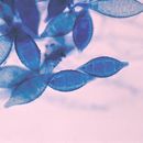
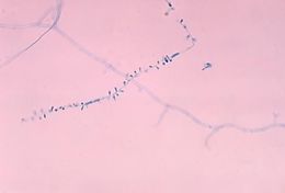
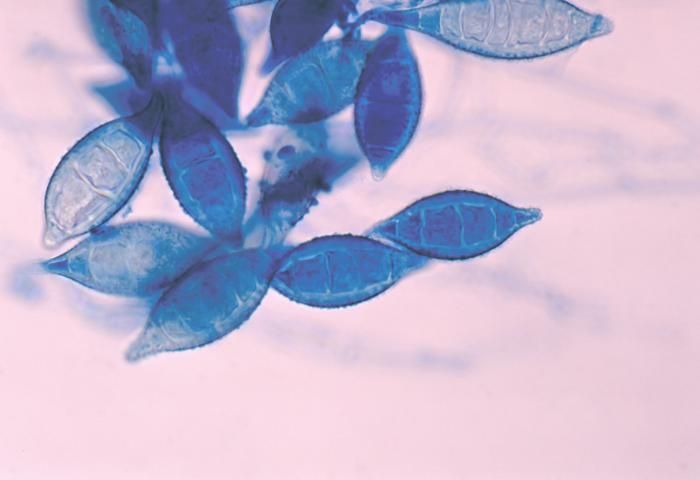
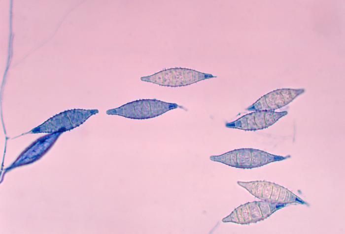
This photomicrograph shows a number of Arthroderma otae, formerly Nannizzia otae, fungal macroconidia.Created: 1978
-
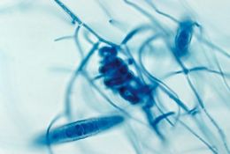
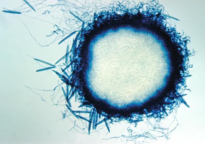
This photomicrograph shows the cleistothecium of the fungus Arthroderma grubyi, formerly Nannizzia grubyia.Created: 1961
-
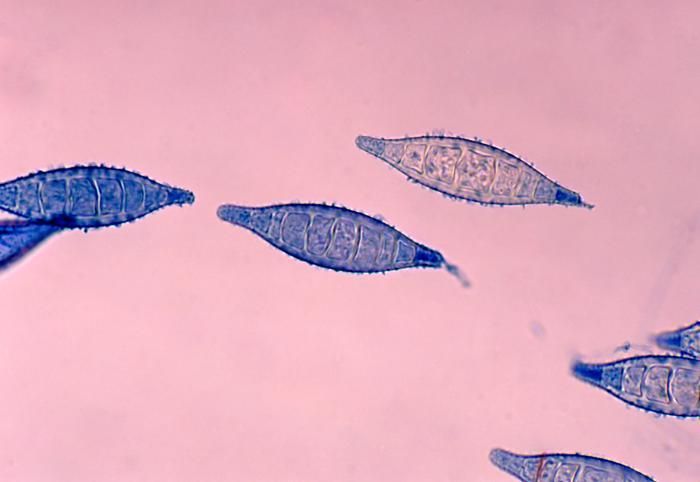
This photomicrograph shows a number of Arthroderma otae, formerly Nannizzia otae, fungal macroconidia.Created: 1978
-
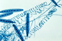
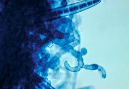
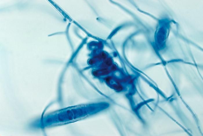
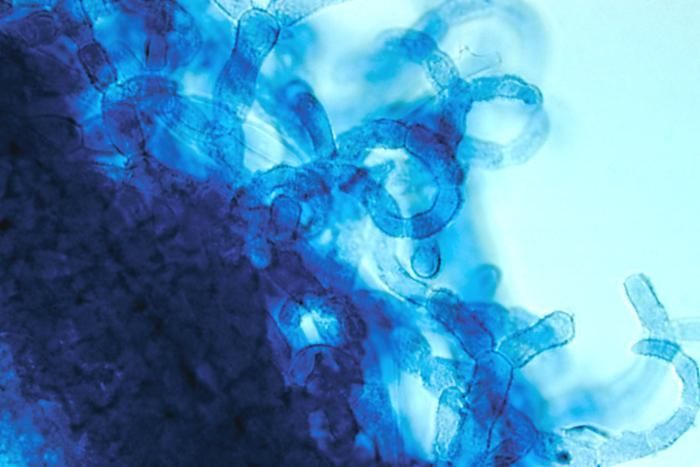
This micrograph reveals spirals and macroconidia from the edge of an immature cleistothecium of the A. grubyi fungus.Created: 1961
-
This photomicrograph shows a number of Arthroderma otae, formerly Nannizzia otae, fungal macroconidia.Created: 1978
-
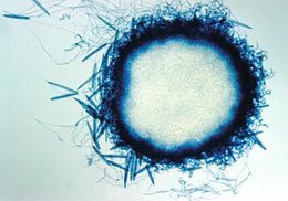
This photomicrograph shows a mature cleistothecium of the fungus Arthroderma grubyi, formerly Nannizzia grubyia.Created: 1961
-
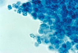
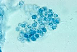
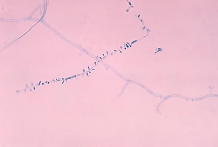
This photomicrograph shows a number of microconidia of the fungus Arthroderma otae, formerly Nannizzia otae.Created: 1978
-
This is the edge of a mature cleistothecium of the fungus Arthroderma grubyi, formerly Nannizzia grubyia.Created: 1961
-
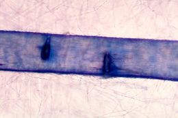
This micrograph reveals the hair perforation caused by the fungus Arthroderma otae, formerly Nannizzia otae.Created: 1978
-
This image shows the cleistothecium of the fungus Arthroderma grubyi, formerly known as Nannizzia grubyia.Created: 1961
-
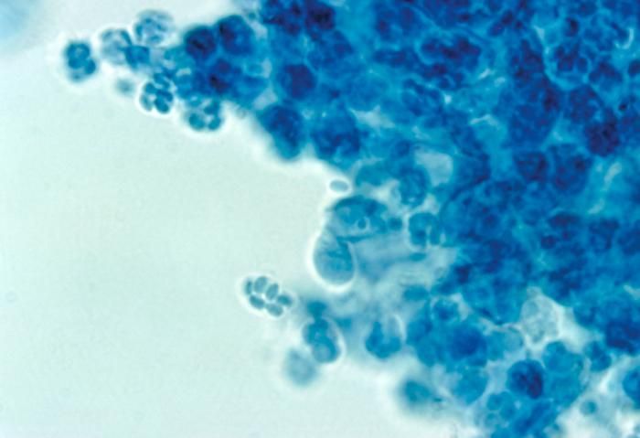
This photomicrograph shows the asci and ascospores of the fungus Arthroderma grubyi, formerly Nannizzia grubyia.Created: 1961
-
This photomicrograph shows the asci and ascospores of the fungus Arthroderma grubyi, formerly Nannizzia grubyia.Created: 1961
-
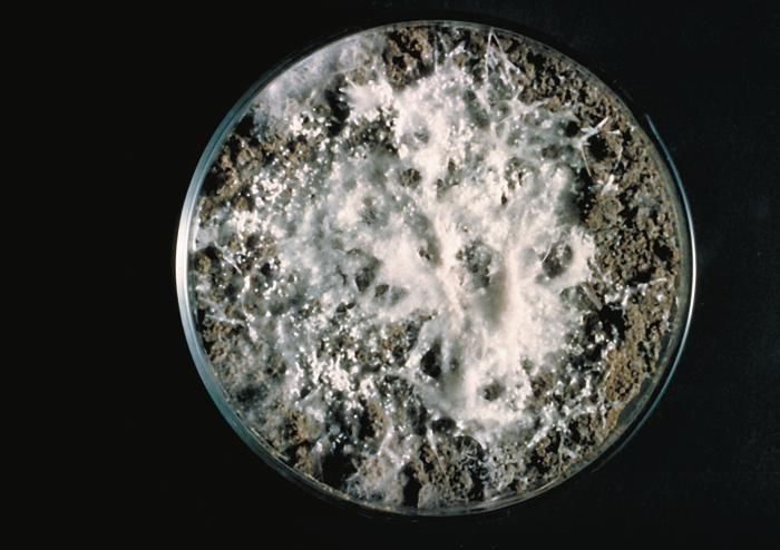
These Arthroderma grubyi cleistothecia were grown on a soil and hair plate culture; formerly Nannizzia grubyia.Created: 1961