Description
provided by Zookeys
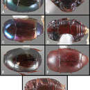
Size range: Length 1.0–5.0mm; width 0.6–4.0mm; Body: ovoid to elongate, sides broadly rounded to sub- or fully parallel, convex to very strongly flattened; color rufescent to frequently piceous or metallic; glabrous or rarely finely setose. Head: frons convex, flat, or deeply depressed, frontal stria usually present along inner margin of eyes, variably interrupted or obsolete across front, frons and epistoma frequently separated by weak to strong transverse carina; supraorbital stria present or absent; epistoma depressed to flat or convex, frequently swollen along apical margin, apical margin usually straight; labrum usually much wider than long, up to 4× or more, usually emarginate apically, but may be straight, bisinuate, or weakly produced; antennal scape usually short, stout, only weakly expanded to apex (Fig. 1A), may be longer, and/or expanded apically; antennal club generally completely tomentose, though rarely glabrous basally, annuli absent, but with 4 characteristic sensory slits on upper and lower surfaces (Fig. 1B), rarely with additional subapical sensorial patch (Fig. 54E); submentum angulate at base, truncate to projecting along distal margin, with few simple setae; gular sutures finely impressed, extending anterolaterad, uninterrupted to basal corner of buccal cavity; mentum subquadrate, sides weakly convergent, apical margin truncate to weakly emarginate, bearing few simple setae; labium with palpifers prominent, palpi with three palpomeres, the basalmost very short, the distal two with short, scattered setae; maxilla with cardo short, transverse, glabrous, stipes triangular, bearing few simple setae, palpi with four palpomeres, the basalmost very short; mandibles (Figs 1A, 3A, 28C) generally each with basal tooth, may be blunt or strong and acute, mandible frequently furrowed along lower, outer edge, may have ventral (mesal) pore and associated (presumed) secretory channel (Fig. 1C). Pronotum: sides parallel to convergent apically; marginal stria usually present and continuous around lateral and anterior margins; lateral submarginal stria present or absent; anterior corners nearly always weakly depressed (Fig. 1D); prescutellar impression absent; disk with single pair of anterior marginal gland openings, usually located close to anterior margin, behind eye on each side, discal punctation highly varied. Elytra: with 2–3 epipleural striae, outer subhumeral stria rarely present, inner subhumeral stria frequently present, often restricted to short basal fragment, dorsal striae 1-5 and sutural stria highly varied, variously abbreviated from base or apex or entirely absent; elytral disk nearly always with distinct secondary punctures in apical half or less (Fig. 1E). Prosternum: prosternal keel varied in width, often quite broad in depressed species, basal margin varied from emarginate to truncate, rarely outwardly arcuate; carinal striae generally present, usually complete, free, rarely abbreviated anteriorly, united or obsolete; prosternal lobe short to moderate in length, apical margin subtruncate to broadly arcuate, rarely bisinuate; marginal stria of prosternal lobe usually distinct across middle, variably obsolete at sides. Mesoventrite: anterior mesoventral margin ranging from distinctly emarginate to distinctly projecting, marginal stria complete to absent, rarely with secondary submarginal stria; mesometaventral stria usually present, most frequently arched forward onto mesoventrite, may in some cases partially displace or completely replace marginal mesoventral stria. Metaventrite: Anterior margin, i.e., mesometaventral suture, frequently arched forward (mirrored in most, but not all cases, in mesometaventral stria), inner lateral metaventral stria generally present, extending from near inner corner of mesocoxa toward metacoxa, or toward posterior corner of metepisternum in the most depressed species, variably sinuate or abbreviated apically; outer lateral metaventral stria present or absent; metaventral disk usually coarsely punctate at sides, impunctate at middle. Abdomen: 1st abdominal ventrite with one or two lateral striae along inner edge of metacoxa, disk usually impunctate at middle, but with conspicuous median punctures in various species; abdominal ventrites 2-5 usually punctate at sides, rarely with dense punctures extending across middle of disk (Fig. 43F). Propygidium often with basal transverse stria (Fig. 5E), disk variably punctate sexually dimorphic in one species; single pair of propygidial gland openings usually conspicuous (Fig. 1F), situated on each side variably near basal margin; pygidium never with apical marginal stria, usually densely punctate, very rarely sexually dimorphic, with male’s setose or otherwise modified; both sexes bearing pygidial trichomes in one species.Legs: Protibia usually rather narrow, with 0-5 unevenly spaced marginal teeth (Fig. 2A), the outer margin nearly always finely serrulate along entire length (Fig. 2A); protibial spurs present, usually short, weakly curved; mesotibia usually with 1-2 weak marginal spines (Fig. 2B), rarely lacking spines; metatibia rarely with any marginal spines (Fig. 2C), generally smooth, with outer apical corner slightly prolonged; tarsi not obviously dimorphic, tarsomeres 1-4 short, usually bearing only single pair of apical setae, tarsomere 5 about as long as 2-4 together, usually weakly dorsoventrally curved (Fig. 2D); tarsal claws simple, separate. Male genitalia:Accessory sclerites absent. T8 generally short, broad, with basal rim strongly sclerotized, basolateral edge extending beneath to inner corner of ventrolateral apodeme; ventrolateral apodeme usually acute, with distal portion strongly reduced, T8 usually broadly open beneath; basal emargination very shallow to moderately deep; basal membrane attachment line rarely evident, usually intersecting basal emargination; distal margin weakly sclerotized, poorly defined, usually vaguely emarginate. S8 articulated at basal corners with ventrolateral apodemes of T8, apical guides weakly developed, halves separate or fused; if separate, halves usually strongly divergent from base, with apices narrow, often rounded, bare to conspicuously setose; if halves of S8 fused, apical margin usually weakly emarginate, frequently with apicoventral velar membrane, bare to conspicuously setose. T9 usually divided, rarely united, with basal apodemes long and slender to short and broad; ventrolateral apodemes weak to strong, opposing or recurved basad, very rarely fused beneath; distal apices usually weakly opposed, subacute to truncate; subapical seta often present on sides. T10 entire, weakly sclerotized, apical margin rounded to weakly emarginate. S9 usually desclerotized along midline, rarely entirely divided; stem very narrow to moderately broad, frequently with ventral keel along much of length; stem rarely absent, with entire T9 short, subcordate; head of S9 usually broad, acute apicolaterally, with apical margin shallowly emarginate to sinuate; tegmen relatively simple, shallowly incised apically, parallel-sided to tapered apically, lacking medioventral tooth or process; median lobe narrow, simple, in a few species associated with small, articulated apical denticulate plates (Fig. 41O); basal piece usually short, with superficial membrane attachment line and oval, asymmetrical basal foramen. Female genitalia: T8 forming a single plate, apical margin usually emarginate; S8 entire or with median plate isolated, with basal baculi detached, articulated with sternites, basally subparallel; S9 usually present, elongate; median coxite articulation present; valviferae paddle-shaped; coxites varied in shape, subquadrate to elongate, with 2-5 apical marginal teeth, with distinct, articulated apical stylus; bursa copulatrix usually completely membraneous, rarely with small sclerotizations of bursal wall; generally with single, bulbous, weakly sclerotized spermatheca, inserted near or at apex of bursa copulatrix; single, basically thin and elongate spermathecal gland present, generally attached near midpoint of spermatheca.
- license
- cc-by-3.0
- copyright
- Michael S. Caterino, Alexey K. Tishechkin
- bibliographic citation
- Caterino M, Tishechkin A (2013) A systematic revision of Baconia Lewis (Coleoptera, Histeridae, Exosternini) ZooKeys 343: 1–297
- author
- Michael S. Caterino
- author
- Alexey K. Tishechkin