-
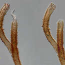
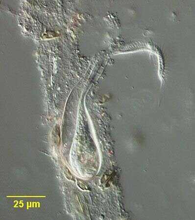
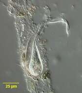
Portrait of the stichotrichine ciliate, Chaetospira remex (Hudson,1875; Kahl,1932). This species occupies a long, sometimes branched tubular lorica into which it intermittently retracts (as seen in this image). The lorica is attached to the substratum. C. muelleri has a flask-shaped lorica. The cell body is slender,elongate and very contractile. The corkscrew shaped anterior bears a prominent adoral zone of membranelles along the peristome. The somatic ciliature is reduced to right and left marginal and two ventral files of short cirri which spiral down the body. The macronucleus is bipartite. the contractile vacuole is in mid-body between the two macronuclei. Feeds mainly on bacteria, flagellates and diatoms. Collected from a freshwater pond near Boise, Idah May 2004. DIC optics.
-
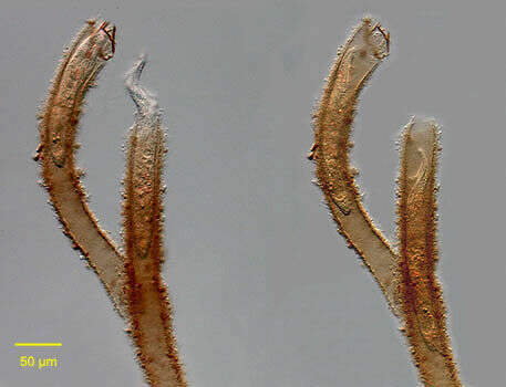
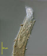
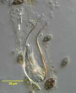
Portrait of the stichotrichine ciliate, Chaetospira remex (Hudson,1875; Kahl,1932). Slightly squashed. This species occupies a long, sometimes branched tubular lorica into which it intermittently retracts. The lorica is attached to the substratum. C. muelleri has a flask-shaped lorica. The cell body is slender,elongate and very contractile. The corkscrew shaped anterior bears a prominent adoral zone of membranelles along the peristome (seen well here). The somatic ciliature is reduced to right and left marginal and two ventral files of short cirri which spiral down the body. The left marginal and one of the ventral cirral files are seen here.The macronucleus is bipartite (not visible in this image). the contractile vacuole is in mid-body between the two macronuclei. Feeds mainly on bacteria, flagellates and diatoms. Collected from a freshwater pond near Boise, Idah May 2004. DIC optics.
-
-
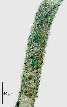
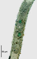
The stichotrichine ciliate, Chaetospira remex (Hudson,1875; Kahl,1932) stained with methyl green-pyronin to demonstrate the bipartite macronucleus. The animal is completely withdrawn into the tubular lorica. The two round macronuclei are stained green. The micronucleus is not seen in this image. The illustration in Kahl's compendium incorrectly depicts C. remex with a single macronucleus (Kahl,A.; Die Tierwelt Deutschlands und der angrenzenden Meeresteile. Teil 25 [Urtiere oder Protozoa I: Wimpertiere oder Ciliata (Infusoria) 3. Spirotricha. Germany:Verlag von Gustav Fischer. 1932, p. 542).Collected from a freshwater pond near Boise Idaho May 2004. Brightfield illumination with closed condenser.
-
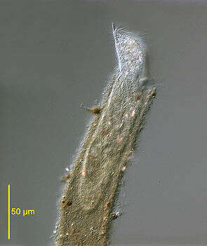
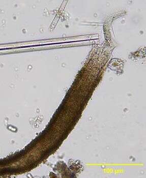
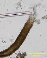
Portrait of Chaetospira, a loricate hypotrich ciliate. Elongate anterior end with prominent adoral zone of membranelles has a corkscrew configuration, distinguishing this genus from the similar Stichotricha. Usually only anterior portion protrudes and organism quickly retracts completely into lorica when disturbed. From freshwater pond near Boise, Idaho. Brightfield.
-
Chaetospira muelleri (LACHMAN,1856). The corkscrew shaped highly contractile anterior end of the cell is seen protriding through the opening of the flask-shaped lorica. Collected from tidal pools at Alki Beach, Seattle, Washington 47°35â41.25âN 122°23â19.60âW.January,2006. DIC.
-
Chaetospira muelleri (LACHMAN,1856). This individual is completely retracted into the the flask-shaped lorica. Collected from tidal pools at Alki Beach, Seattle, Washington 47°35â41.25âN 122°23â19.60âW.January,2006. DIC.