-
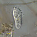
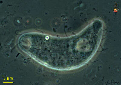
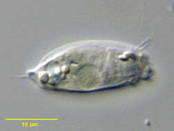
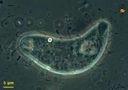
Portrait of Rhopalophrya gracilis (Kahl,1926).Collected from a freshwater pond near Boise, Idaho. DIC.
-
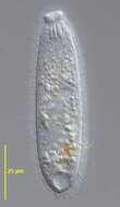
Enchelys (ench-el-is), anterior end of this predatory ciliate, picture taken to show the rows of cilia or kineties which extend from the front of the cell to the posterior. Phase contrast.
-
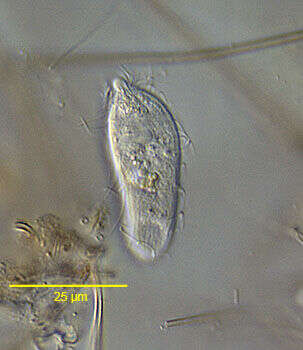
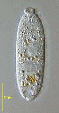
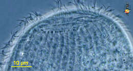
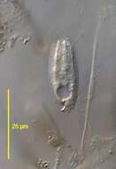
Surface view of Pithothorax processus a small haptorid ciliate found in polysaprobic habitats. The body is a slightly flattened cylinder. The pellicle is rigid with longitudinal ribbing. The oral aperture is at the anterior apex surrounded by projections of the pellicular ridges. There is a curved funnel-shaped posterior process from which a long caudal cilium protrudes (seen here). The round macronucleus is anterior and the contractile vacuole is located in the posterior 1/3 at the periphery (seen here). The somatic ciliature is confined to the anterior and posterior quarters of the body. From stagnant organically enriched freshwater pond near Boise, Idaho. DIC optics.
-
Portrait of Rhopalophrya gracilis (Kahl,1926).Collected from a freshwater pond near Boise, Idaho. DIC.
-
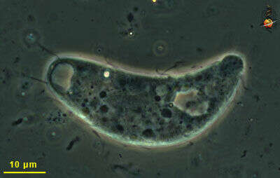
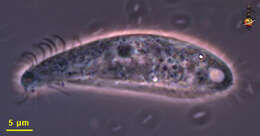
Enchelys (ench-el-is) -cylindrical predatory ciliate, body fairly flexible, mouth slit zone at anterior end, underlain by a number of extrusomes. The cell has just eaten a dinoflagellate. Differential interference contrast.
-
Saggital optical section of Pithothorax processus (Kahl, 1926), a small haptorid ciliate found in polysaprobic habitats. The body is a slightly flattened cylinder. The pellicle is rigid with longitudinal ribbing. The oral aperture is at the anterior apex surrounded by projections of the pellicular ridges. There is a curved funnel-shaped posterior process from which a long caudal cilium protrudes (seen here). The round macronucleus is anterior and the contractile vacuole is located in the posterior 1/3 at the periphery (seen here). The somatic ciliature is confined to the anterior and posterior quarters of the body. From stagnant organically enriched freshwater pond near Boise, Idaho. DIC optics.
-
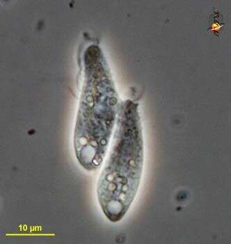
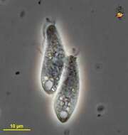
Enchelys (en-chill-iss) small predatory ciliate, here two cells are linked in conjugation (during which time DNA is exchanged - it+s a kind of sexual activity but without reproduction). Clear vacuoles at the rear are contractile vacuoles. Phase contrast micrograph.
-
Surface view of Pithothorax processus (Kahl, 1926) a small haptorid ciliate found in polysaprobic habitats. The body is a slightly flattened cylinder. The pellicle is rigid with longitudinal ribbing. The oral aperture is at the anterior apex surrounded by projections of the pellicular ridges. There is a curved funnel-shaped posterior process from which a long caudal cilium protrudes (seen here). The round macronucleus is anterior and the contractile vacuole is located in the posterior 1/3 at the periphery (seen here). The somatic ciliature is confined to the anterior and posterior quarters of the body. From stagnant organically enriched freshwater pond near Boise, Idaho. DIC optics.
-
Enchelydium KAHL,1930, species undetermined.Phase contrast.
-
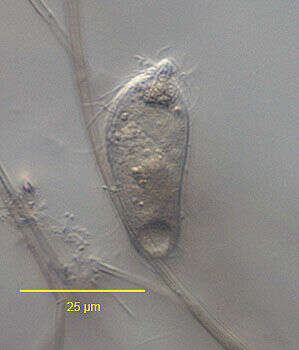
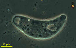
In vivo portrait of Pithothorax processus (Kahl, 1926), a small haptorid ciliate found in polysaprobic habitats. The body is a slightly flattened cylinder. The pellicle is rigid with longitudinal ribbing. The oral aperture is at the anterior apex surrounded by projections of the pellicular ridges. There is a curved funnel-shaped posterior process from which a long caudal cilium protrudes (seen here). The round macronucleus is anterior and the contractile vacuole is located in the posterior 1/3 at the periphery (seen here). The somatic ciliature is confined to the anterior and posterior quarters of the body. From stagnant organically enriched freshwater pond near Boise, Idaho. June 2005. DIC optics.
-
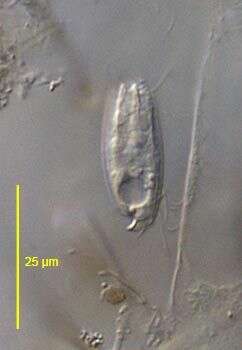
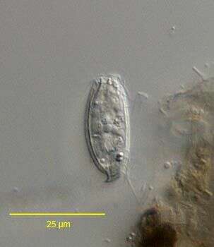
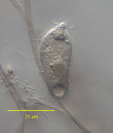
Small ciliate, anterior region of cell is to the right. With a small number of kineties, and with cilia sparsely distributed. We are uncertain of the identity of this cell. Phase contrast micrograph.
-
Portrait of Pithothorax processus (Kahl, 1926), a small haptorid ciliate found in polysaprobic habitats. The body is a slightly flattened cylinder. The pellicle is rigid with longitudinal ribbing. The oral aperture is at the anterior apex surrounded by pointed projections of the pellicular ridges. There is a funnel shaped posterior process from which a long caudal cilium protrudes (visible here). The round macronucleus is anterior. The somatic ciliature is confined to the anterior and posterior quarters of the body. Although this specimen was stained by the silvercarbonate technic (see Foissner, W. Europ. J. Protistol., 27:313-330;1991, the somatic kinetids did not impregnate. Short rod shaped partially discharged extrusomes (stained brown here) are visible anteriorly. From stagnant organically enriched freshwater pond near Boise, Idaho. Brighfield.
-
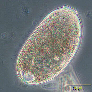
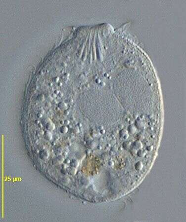
A very small ciliate, the anterior end of which has protruded. There is a contractile vacuole at the posterior end and sparse ciliation. We are unsure of the identity of this organism. Phase contrast micrograph.
-
In vivo portrait of Pithothorax processus (Kahl, 1926), a small haptorid ciliate found in polysaprobic habitats. The body is a slightly flattened cylinder. The pellicle is rigid with longitudinal ribbing. The oral aperture is at the anterior apex surrounded by projections of the pellicular ridges. There is a curved funnel-shaped posterior process from which a long caudal cilium protrudes (seen here). The round macronucleus is anterior and the contractile vacuole is located in the posterior 1/3 at the periphery (seen here). The somatic ciliature is confined to the anterior and posterior quarters of the body. From stagnant organically enriched freshwater pond near Boise, Idaho. June 2005. DIC optics.
-
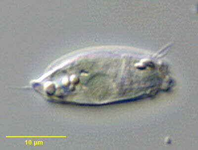
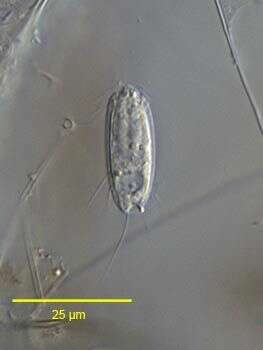
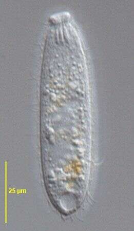
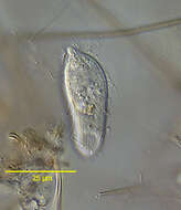
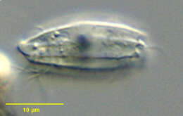
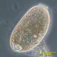
This small predatory ciliate has its mouth at the anterior end (to the left). The body is evenly ciliated but there are some longer stiff cilia near the mouth. The contractile vacuole is the light area near the end of the cell. Phase contrast microscopy.
-
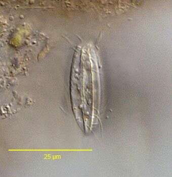
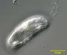
Cross-sectional view of Pithothorax processus (Kahl, 1926) a small haptorid ciliate found in polysaprobic habitats. The body is a slightly flattened cylinder. The pellicle is rigid with longitudinal ribbing giving it a fluted appearance. From stagnant organically enriched freshwater pond near Boise, Idaho.June 2005. DIC.
-
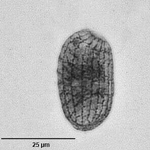
Enchelydium fusidens KAHL, 1930. This population is about 25% smaller than the one described by KAHL.The important morphological featues (oral bulge, extrusomomes, macronucleus) match those of E. fusidens. DIC.
-
Lasteral view of Pithothorax processus (Kahl, 1926), a small haptorid ciliate found in polysaprobic habitats. The body is a slightly flattened cylinder. The pellicle is rigid with longitudinal ribbing. The oral aperture is at the anterior apex surrounded by projections of the pellicular ridges. There is a curved funnel-shaped posterior process from which a long caudal cilium protrudes. The round macronucleus is anterior and the contractile vacuole is located in the posterior 1/3 at the periphery. The somatic ciliature is confined to the anterior and posterior quarters of the body. From stagnant organically enriched freshwater pond near Boise, Idaho. June 2005. DIC optics.
-
Fully contracted Enchelydium fusidens KAHL, 1930. This population is about 25% smaller than the one described by Kahl.The important morphological featues (oral bulge, extrusomomes, macronucleus) match those of E. fusidens. DIC.
-
partially contracted Enchelydium fusidens KAHL, 1930. This population is about 25% smaller than the one described by Kahl.The important morphological featues (oral bulge, extrusomomes, macronucleus) match those of E. fusidens. DIC.
-
Enchelydium fusidens KAHL, 1930. This population is about 25% smaller than the one described by Kahl.The important morphological featues (oral bulge, extrusomomes, macronucleus) match those of E. fusidens. DIC.
-
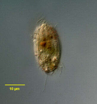
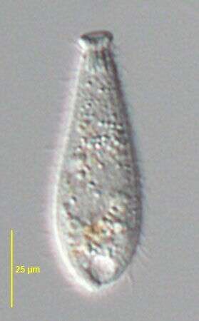
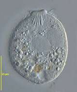
Portrait of the small Enchelyid ciliate, Pithothorax rotundus (Kahl, 1927). The body is cylindrical, bluntly tapered anteriorly and rounded posteriorly. The right side is gently convex, the left side more straight giving the anterior portion the appearance of being bent slightly to the left. The pellicle is rigid with longitudinal ridges much less prominent than those seen in P. processus. The ridges converge around the anterior apical cytostome. The cytopharynx is supported by delicate trichites. In this image a cluster of discharged extrusomes is seen to the viewer's right. The somatic cilia are reduced to a few kineties in the anterior and posterior quarter of the cell. The macronucleus is central and the single contractile vacuole posterior. Pithothorax is a sapropelic genus usually found in hypoxic bottom sediments with decomposing organic matter. Collected from bottom sediments of freshwater aquaculture tank near Boise, Idaho January 2004. DIC optics.
-
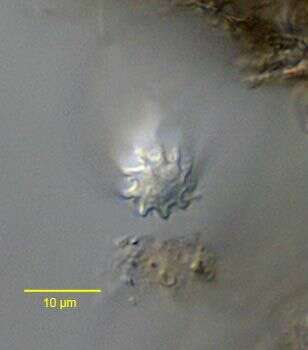
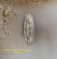
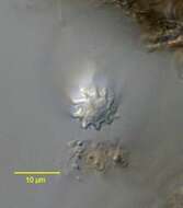
Silverline system (ventral view) of Pithothorax rotundus (Kahl,1927). Collected from sapropelic bottom sediments of a slow-moving freshwater stream near Boise, Idaho. Stained by the dry silver nitrate technic (see Foissner, W.Europ. J. Protistol.27,313-330;1991). Brightfield.