Nan-Nan Li, Masanori J. Toda, Zhao Fu, Ji-Min Chen, Su-Hua Li, Jian-Jun Gao
Zookeys
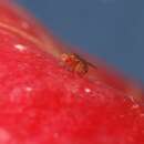
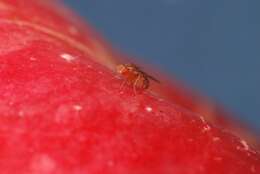
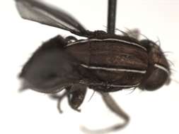
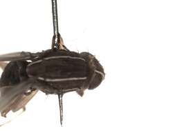
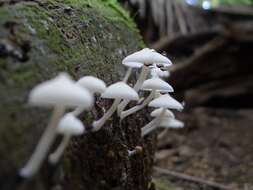
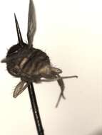
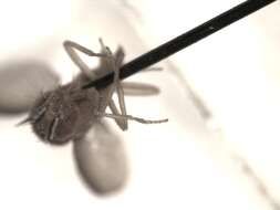
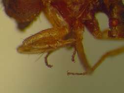
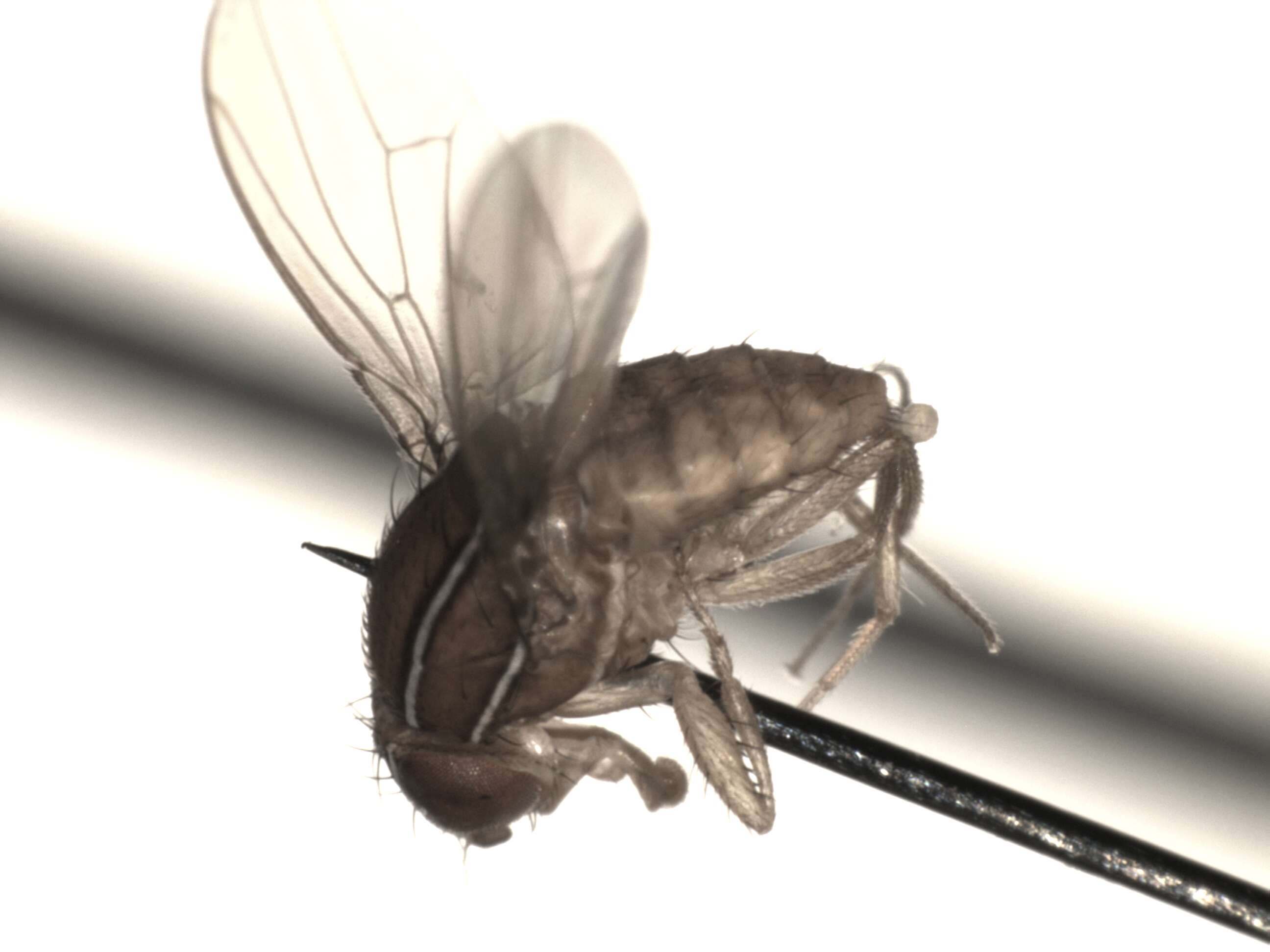
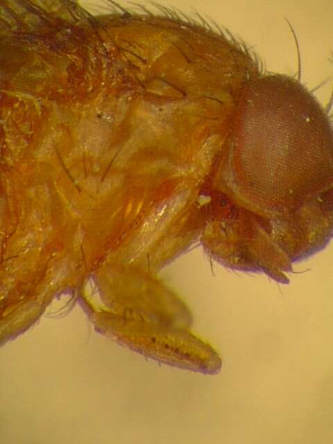
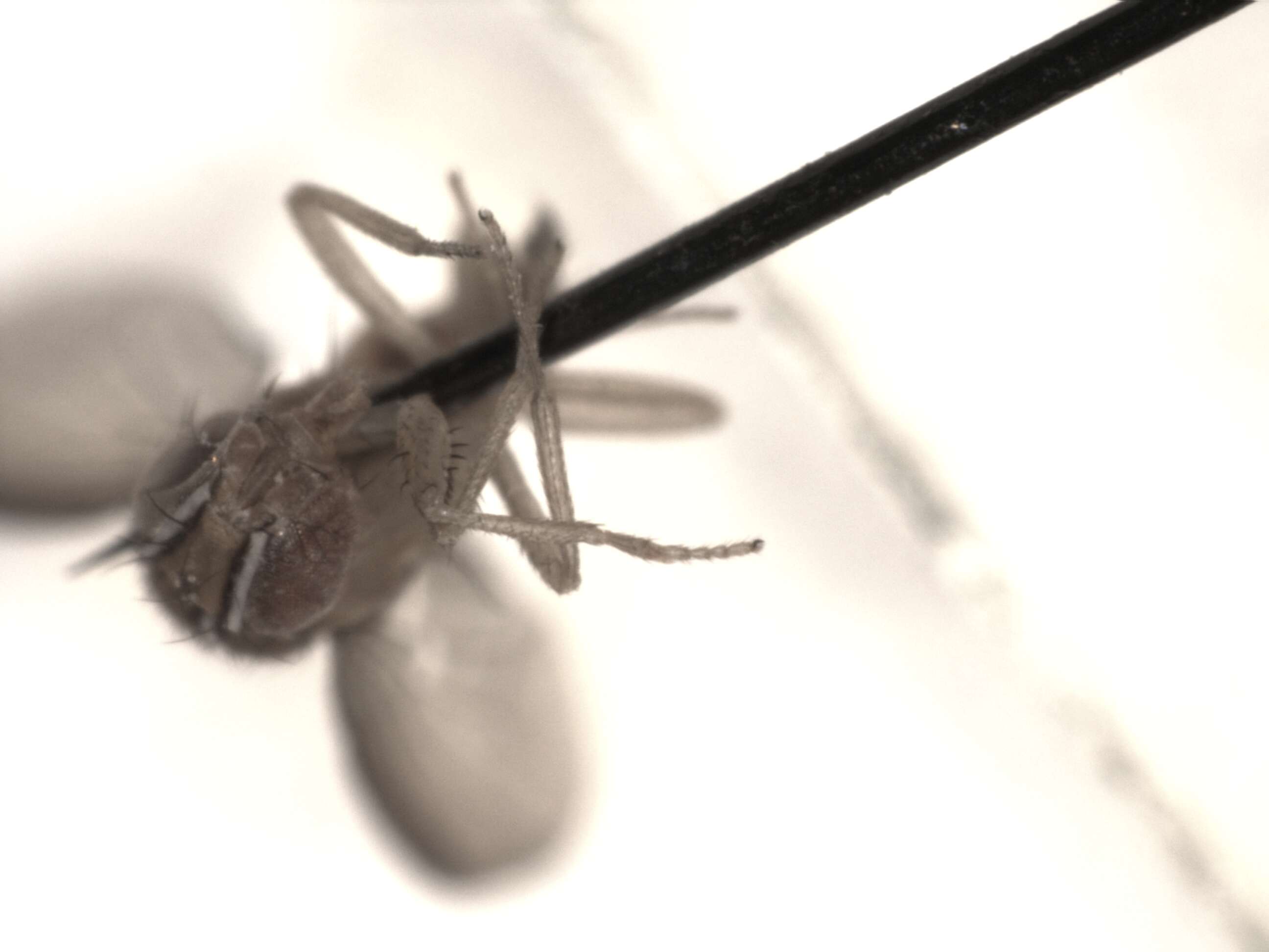
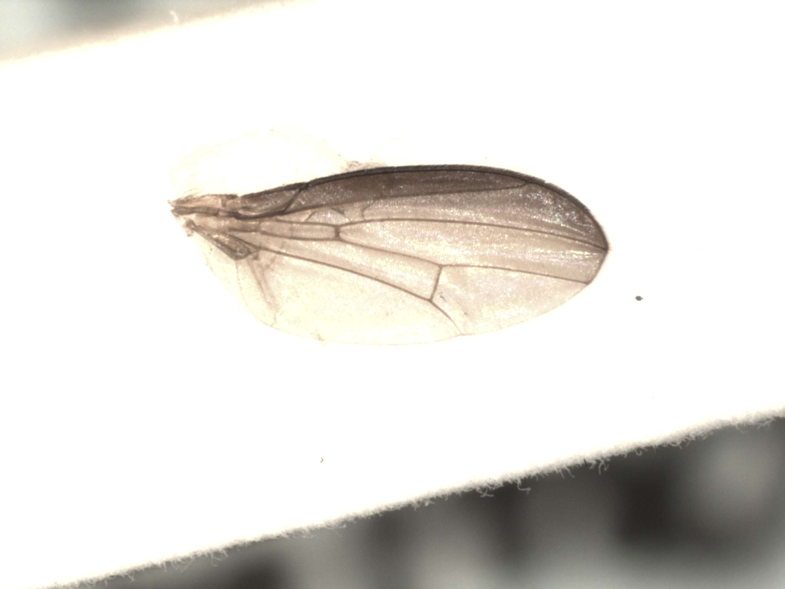
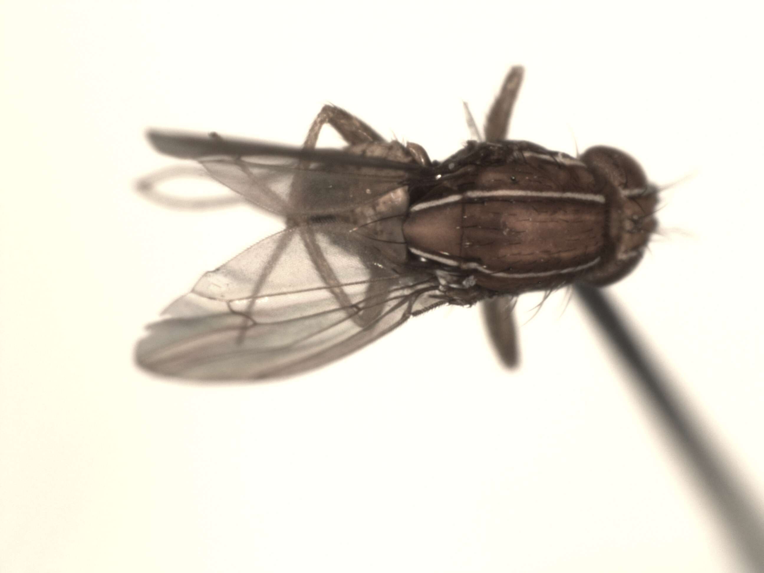
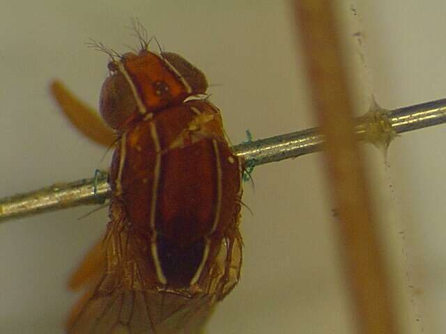
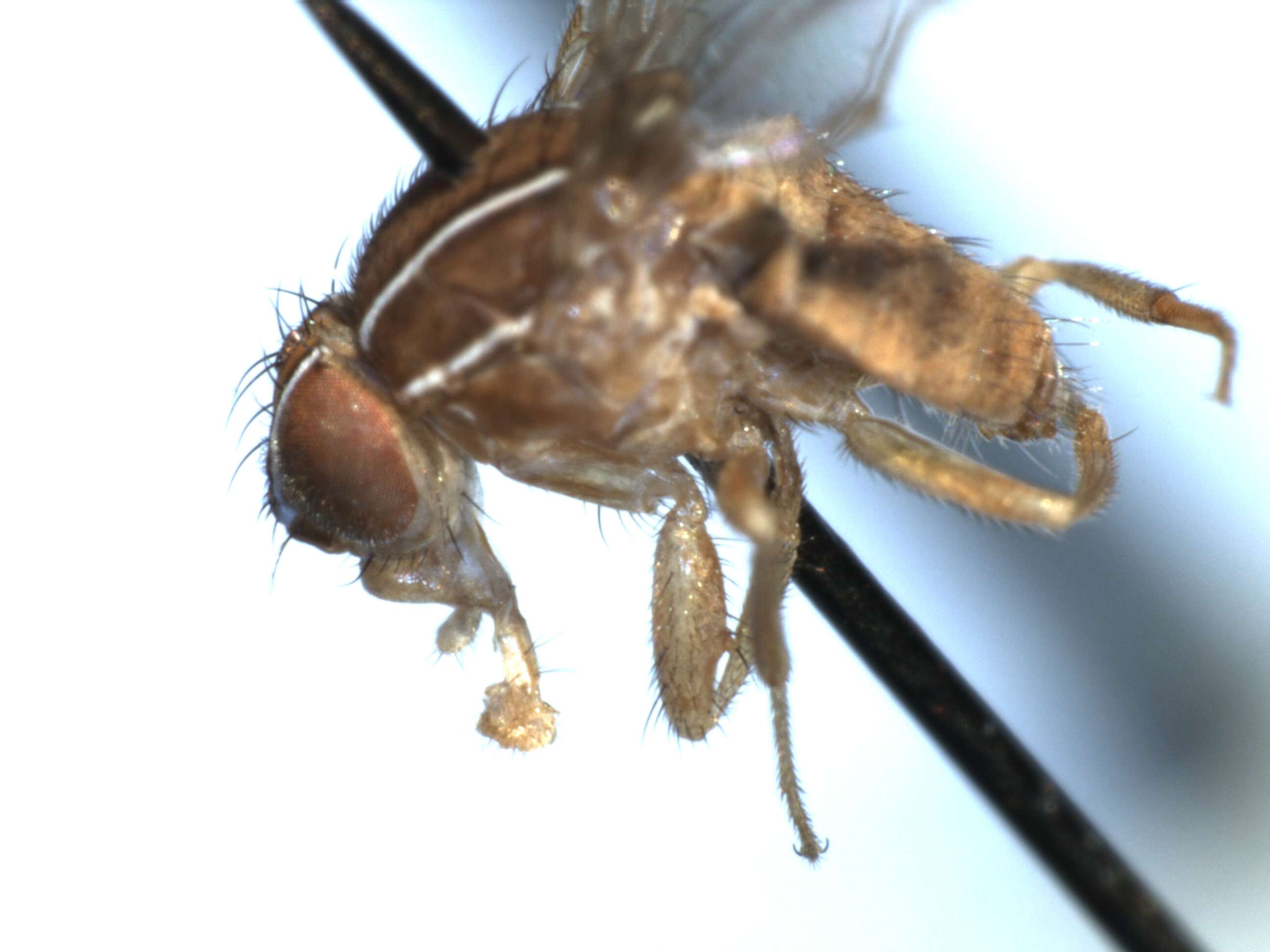
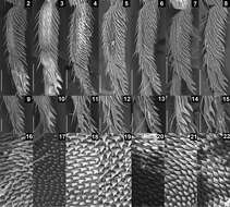
Figures 2–22.SEM photographs showing leg and oviscapt fine structures in the Colocasiomyia gigantea species group. Foreleg tarsomeres I and II (2–8), pegs on foreleg tarsomere II (9–15) and warts on basal part of lateral lobe or basal membrane of oviscapt (16–22) of Colocasiomyia gigantea (2, 9, 16), Colocasiomyia scindapsae (3, 10, 17), Colocasiomyia rhaphidophorae (4, 11, 18), Colocasiomyia longifilamentata sp. n. (5, 12, 19), Colocasiomyia longivalva sp. n. (6, 13, 20), Colocasiomyia hailini sp. n. (7, 14, 21) and Colocasiomyia yini sp. n. (8, 15, 22). Scale line = 0.1 mm in 2–8, 0.05 mm in 9–15. Figures 16–22 are in the same magnification, with the width corresponding to 30 µm.