dcsimg
Image of Brachionus manjavacas Fontaneto, Giordani, Melone & Serra 2007
Creatures » » Animal » » Rotifers » » Brachionidae »

Brachionus manjavacas Fontaneto, Giordani, Melone & Serra 2007

Behaviour

provided by Marine Rotifera LifeDesk

Monogonont rotifers, which include Brachionus, are cyclical parthenogens; they proliferate asexually in the laboratory forming clones, with males being much more infrequent than females. Brachionus manjavacas is a planktonic rotifer moving by cilia-beating present on the head.

license
cc-by-nc
copyright
Leasi, Francesca
author
Leasi, Francesca

Biology

provided by Marine Rotifera LifeDesk

Brachionus manjavacas lives in brackish water habitats, including inland salt waters.This species is mainly parthenogenetic with the occasional generation of males. Sexual reproduction produces diapausing embryos (resting eggs).

Type locality: Laguna de Manjavacas, Guadiana basin, Spain.


license
cc-by-nc
copyright
Leasi, Francesca
author
Leasi, Francesca

Description

provided by Marine Rotifera LifeDesk
license
cc-by-nc
copyright
Leasi, Francesca
author
Leasi, Francesca

Diagnostic Description

provided by Marine Rotifera LifeDesk

Brachionus manjavacas is a species of the Ltype B. plicatilis complex, and cannot be reliably distinguished from B. plicatilis s.s. on lorica shape, body size, shape of anterior spines, and trophi size, so that a morphological stasis has been proposed and here confirmed, despite ancient divergence. Presently, the only available morphological feature to distinguish B. manjavacas from B. plicatilis s.s. is the shape of the satellites of the rami, which are much more acutely pointed along the inner upper margin in B. manjavacas. The most reliable feature to discriminate this species is still molecular barcoding, and genbank deposited sequences for this clone are AF387257 for COI, and AF387213 for ITS1 for SEM, in the collection of G. Melone at the University of Milan, Department of Biology.

Type locality: Laguna de Manjavacas, Guadiana basin, Spain, approx. coord. 39'25°N, 2'53'W.


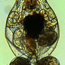
Holotype: A parthenogenetic female in a permanent, glycerin glass slide mount deposited in the Museo civico di Storia Naturale di Milano (MSNM), Italy, catalogue nr. IV75.


Paratypes: Two slides with one specimen each, catalogue MSNM nr. IV76–77; one stub with 20 parthenogenetic females prepared for SEM, and three stubs with 8, 9, and 46 trophi preparations

license
cc-by-nc
copyright
Leasi, Francesca
author
Leasi, Francesca

Distribution

provided by Marine Rotifera LifeDesk

This species has been found in Spain at the Laguna of Manjavacas. It is known from other Spanish sampling sites, other than the type locality: Laguna del Camino de Villafranca, Laguna de Tìrez . Other sites where the species has been found previously are: Spain (Laguna de Gallocanta, Laguna del Longar de Lillo, Laguna de Fuente de Piedra, Laguna de Capacete, Laguna de Pétrola). Tunisia (Korba ebkhet), and Russia (Sea of Azov).

license
cc-by-nc
copyright
Leasi, Francesca
author
Leasi, Francesca

Ecology

provided by Marine Rotifera LifeDesk

In brackish water habitats, including inland salt waters.

license
cc-by-nc
copyright
Leasi, Francesca
author
Leasi, Francesca

Genetics

provided by Marine Rotifera LifeDesk

Genbank deposited sequences for this clone are AF387257 for COI, and AF387213 for ITS1.

license
cc-by-nc
copyright
Leasi, Francesca
author
Leasi, Francesca

Habitat

provided by Marine Rotifera LifeDesk

Saltwater habitat, planktonic.

license
cc-by-nc
copyright
Leasi, Francesca
author
Leasi, Francesca

Life Cycle

provided by Marine Rotifera LifeDesk

Parthenogenesis dominates the life cycle where reproduction occurs in the absence of males (amictic phase). In response to certain environmental cues, such as temperature, population density, and food quality and quantity changes, males may be produced and sexual reproduction takes place (mictic phase)
Amictic females are diploid and produce diploid (amictic) eggs. Mictic females produce male eggs which are smaller and more numerous than amictic eggs. Fertilized mictic females produce a diapausing embryo called a resting egg.

license
cc-by-nc
copyright
Leasi, Francesca
author
Leasi, Francesca

Look Alikes

provided by Marine Rotifera LifeDesk

Brachionus plicatilis Müller, 1786

license
cc-by-nc
copyright
Leasi, Francesca
author
Leasi, Francesca

Migration

provided by Marine Rotifera LifeDesk

Without active dispersal stage: direct development. However, passive and not active dispersing stages exist, during both embryo- (eggs and resting eggs) and adult phase.

license
cc-by-nc
copyright
Leasi, Francesca
author
Leasi, Francesca

Molecular Biology

provided by Marine Rotifera LifeDesk

http://www.ncbi.nlm.nih.gov/Taxonomy/Browser/wwwtax.cgi?id=667381&lvl=0

license
cc-by-nc
copyright
Leasi, Francesca
author
Leasi, Francesca

Morphology

provided by Marine Rotifera LifeDesk

Parthenogenetic female. Lorica soft, shape pear-like, very similar to B. plicatilis. Anterior dorsal margin of the lorica with three pairs of spines flanking the U-shaped sinus; anterior ventral margin with two pairs of rounded lobules. Corona with typical features. Foot aperture sub-terminal; end of foot with two main toes and a smaller median toe. Lateral antennae medially located. Trophi. Malleate type, Fulcrum short, flattened laterally. Rami broad, rounded externally, asymmetrical, usually right ramus longer; inner margins with uniform zig-zag like reinforced ridge; satellites prominent, each with an acute point characteristic of the species. Unci with 6-8 teeth; shafts and teeth almost completely fused; teeth gradually decreasing in size from the ventral one, with the second tooth, either on left or on right uncus, bigger than the first one; distal margin of uncus plate, connecting to manubria, V-shaped, with shortest section of V ventral. Subunci brush-like, with many scleropili. Manubria with crest on dorsal surface of medium height.

MUSCULATURE:

Muscular system of B. manjavacas comprised fibers mainly arranged in longitudinal and circular orientations. Muscles were present as fibers of the body wall (somatic) or present in a visceral (splanchnic) position. Somatic muscles consisted of inner longitudinal and outer dorsoventral arrangements, except for circular muscles of the head (pars coronalis and coronal sphincter) and at the junction of foot and toes (circumpedalis), common in most rotifers. Splanchnic muscles were present in the gut of females and reproductive apparatus of males. Between the two sexes, differences in the general organization of the somatic musculature were not observed, except for the musculature of the head. The muscle pars coronalis was complete in females, but ventrally open in males; the coronal sphincter was present only in males. A few other differences were seen in the diameter and points of insertion of muscular fibers.

For trophi morphology see: http://users.unimi.it/melone/trophi/start.html

license
cc-by-nc
copyright
Leasi, Francesca
author
Leasi, Francesca

Reproduction

provided by Marine Rotifera LifeDesk

Cyclically parthenogenetic and sexual.

license
cc-by-nc
copyright
Leasi, Francesca
author
Leasi, Francesca

Size

provided by Marine Rotifera LifeDesk

Measurements (range, and in parenthesis, mean ± standard error, in µm): Parthenogenetic female (N = 0, optical microscopy). Lorica length 217.5-312.0 (258.0 ± 2.5), lorica width 147.0-205.0 (172.5 ± 2.0).
Trophi. Larger ramus length 23.07-36.34 (28.91 ± 0.28, N = 89), smaller ramus length 21.22-32.55 (26.34 ± 0.28, N = 89), uncus length 16.92-33.49 (26.97 ± 0.16, N = 251), manubrium length 9.65-48.21 (38.53 ± 0.20, N = 376).

license
cc-by-nc
copyright
Leasi, Francesca
author
Leasi, Francesca

Biogeography

provided by Rotifers of the World
Palaearctis
Nearctis
license
cc-by-nc
original
visit source
partner site
Rotifers of the World