-
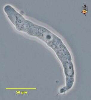
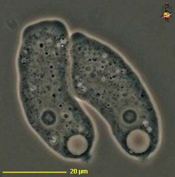
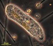
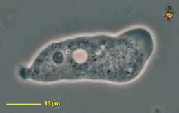
Hartmannella (heart-man-ella), a naked amoeba, limax (slug-like) body form, well developed hyaline cap, central nucleus and scrunched up uroidal region. Phase contrast.
-
-
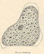
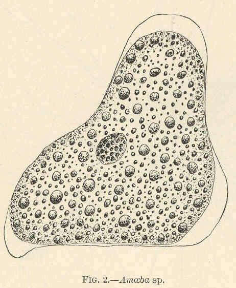
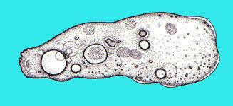
Amoeba sp..
-
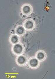
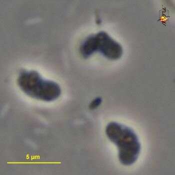
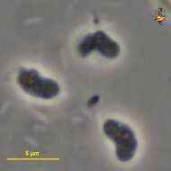
Hartmannella (heart-man-ella), a naked amoeba, limax (slug-like) body form, cysts. Phase contrast.
-
-
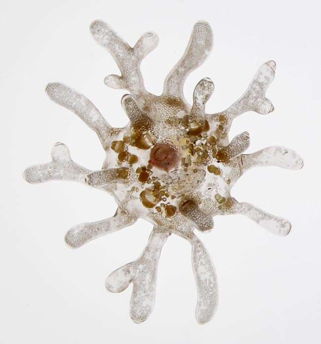
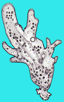
Amoeba roteus
-
Hartmannella (heart-man-ella), a naked amoeba, limax (slug-like) body form, well developed hyaline cap, central nucleus and scrunched up uroidal region. Phase contrast.
-
-
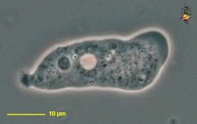
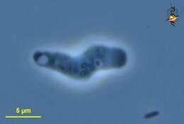
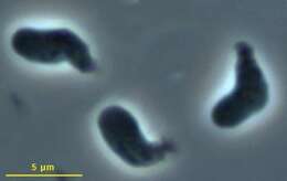
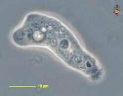
Cashia (cash-ee-a) - tentative identification - small limax (slug-shaped) amoeba, hyaline cap to right lacks inclusions, contractile vacuole is associated with the posterior end of the cell. Phase contrast.
-
-
Cashia. Cell observed in freshwater sediments in the vicinity of Broome, Western Australia in September 2003. This image was taken using phase contrast optics. This work was supported by the Australian Biological Resources Study.
-
Nolandella (no-lane-ell-a) is a small naked amoeba. Phase contrast micrograph.
-
-
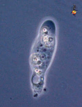
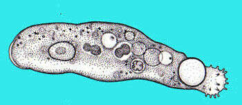
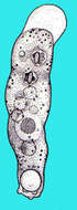
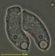
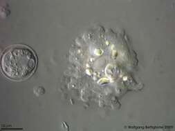
Saccamoeba (sack-a-me-ba), a monopodial naked free-living amoeba. With a lobose pseudopodium, usually progressing as a single pseudopodium (i.e. is monopodial). Hyaline cap absent or not well developed. Small uroid. These cells also with light-coloured contractile vacuoles and with nuclei with nucleoli. Phase contrast.
-
Saccamoeba (sack-a-me-ba), a monopodial naked free-living amoeba. With a lobose pseudopodium, usually progressing as a single pseudopodium (i.e. is monopodial). Hyaline cap absent or not well developed. Small uroid. This cell also with a light-coloured contractile vacuole and nucleus with nucleolus. Phase contrast.
-
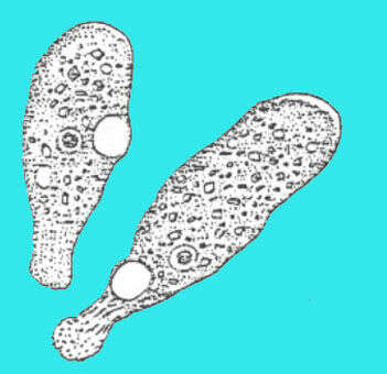
-
-
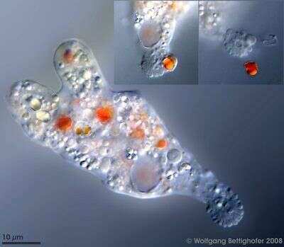
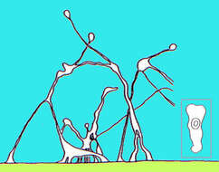
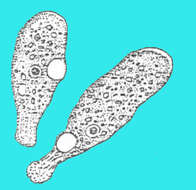
Saccamoeba species with its villous uroid. The two pseudopods protruded during change of direction. During ordinary movement on substrate the appearance is monopodial. Scale bar indicates 10 µm. The inserts show states of defecation. Sample from sphagnum pond Dosenmoor near Neumuenster (Schleswig-Holstein, Germany). This image was taken using Zeiss Universal with Olympus C7070 CCD camera.
-
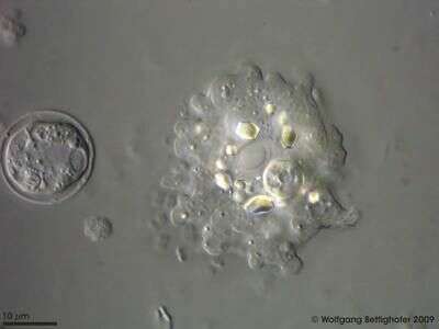
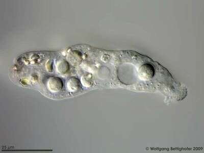
Floating from of Saccamoeba limax with a charcteristic villous collar and the cuboidal crystals. The vesicular nucleus with its huge central nucleolus lies in the middle of the cell. Left of Saccamoeba we can see the prey: Pyxidicula, a testate amoeba. Scale bar indicates 10 µm. Sample from a freshwater pond on the island of Hiddensee (Baltic Sea, Germany). This image was taken using Zeiss Universal with Olympus C7070 CCD camera.
-
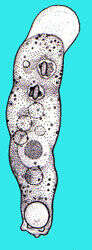
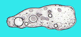
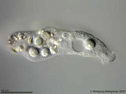
Saccamoeba limax in classical shape as it can be found in literature. From the left to the right we see the truncated bipyramidal crystals (typical for this species), several food vacoules, the vesicular nucleus with its huge central nucleolus, the contractile vacuole and another food vacuole. On the right the likewise typical finely papillate bulbous uroid which resembles the family Amoebidae.Scale bar indicates 25 µm. Sample from a freshwater pond on the island of Hiddensee (Baltic Sea, Germany). This image was taken using Zeiss Universal with Olympus C7070 CCD camera.
-
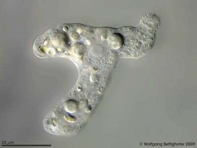
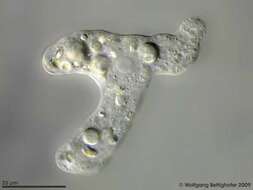
Saccamoeba limax branching and changing its direction. During this period the classical, monopodial shape, is left. Scale bar indicates 25 µm. Sample from a freshwater pond on the island of Hiddensee (Baltic Sea, Germany). This image was taken using Zeiss Universal with Olympus C7070 CCD camera.
-
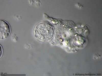
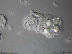
Saccamoeba limax starting the uptake of a Pyxidicula cell. Scale bar indicates 25 µm. Sample from a freshwater pond on the island of Hiddensee (Baltic Sea, Germany). This image was taken using Zeiss Universal with Olympus C7070 CCD camera.
-