-
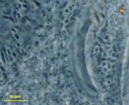
Phase contrast micrograph showing clearly one contractile vacuole with five filled channels.
-
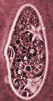
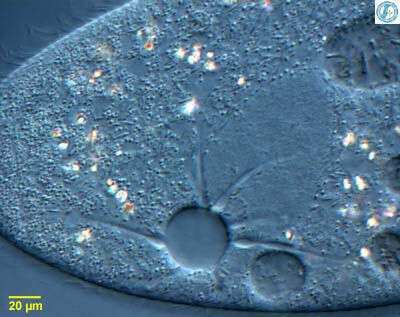
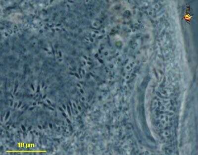
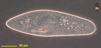
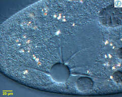
Paramecium with its iconic contractile vacuole. The central vacuole is surrounded by an array of collecting canals which collect fluid from the cytoplasm and feed it into the vacuole. The region of the canals near the vacuole are distensible ampullae. From Lake Donghu, China. Differential interference contrast micrograph.
-
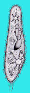
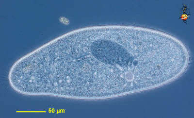
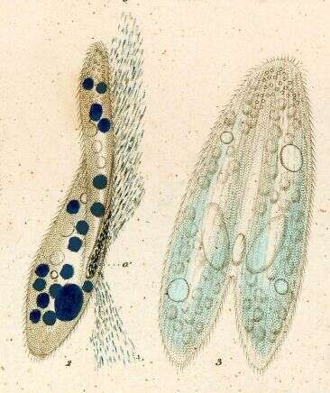
Paramecium (aurelia) (par-a-mee-see-um) is a very familiar genus of ciliates. They eat bacteria and have the mouth recessed in a buccal cavity, and the cell is often shaped with a scoop leading to the mouth. There are cilia all over the body with a caudal tuft of longer cilia at the back of the body. Usually with a layer of extrusomes (trichocysts) under the cell surface and a large oval macronucleus. Contractile vacuoles star-shaped. This species is P. aurelia, one of the smaller spindle-shaped (morpho)species. The (morpho) species is best distinguished by the presence of two small micronuclei pressed up against the macronucleus. Phase contrast.
-
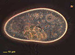
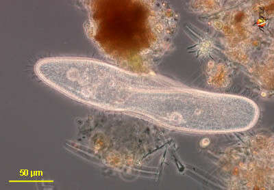
The Paramecium, shown in this image, was flattened slightly to make the cell components more visible. The cell is covered with hundreds of cilia. The dark structure upper right is the macronucleus.
-
Paramecium (aurelia) (par-a-mee-see-um) is a very familiar genus of ciliates and this (morpho) species is best distinguished by the presence of two small micronuclei pressed up against the macronucleus. They can be seen here to the north of the nucleus. Differential interference contrast.
-
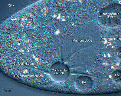
Paramecium with its iconic contractile vacuole. The central vacuole is surrounded by an array of collecting canals which collect fluid from the cytoplasm and feed it into the vacuole. The region of the canals near the vacuole are distensible ampullae. This image has been annotated by Marc Perkins at Orange Coast College (a community college in Costa Mesa, California).
-
Paramecium (aurelia) (par-a-mee-see-um) is a very familiar genus of ciliates and this (morpho) species is best distinguished by the presence of two small micronuclei pressed up against the macronucleus. This image shows the peniculi or compound ciliary organelles in the mouth. Phase contrast.
-
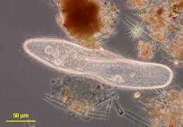
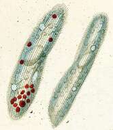
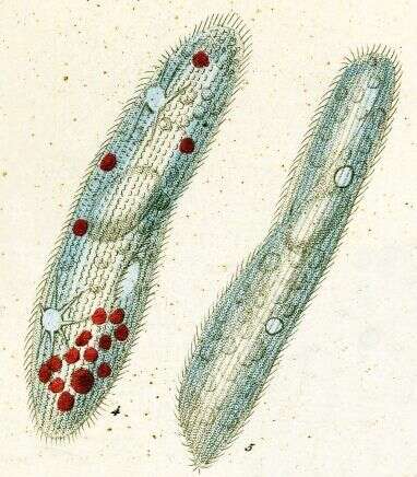
Paramecium (caudatum) (par-a-mee-see-um) is a very familiar genus of ciliates. They eat bacteria and have the mouth recessed in a buccal cavity, and the cell is often shaped with a scoop leading to the mouth. There are cilia all over the body with a caudal tuft of longer cilia at the back of the body. Usually with a layer of extrusomes (trichocysts) under the cell surface and a large oval macronucleus. Contractile vacuoles star-shaped. This species is P. caudatum, and was photographed with the cell pushing itself into some debris. This is the normal feeding behaviour of this genus. Phase contrast.
-
Paramecium (aurelia) (par-a-mee-see-um) is a very familiar genus of ciliates and this (morpho) species is best distinguished by the presence of two small micronuclei pressed up against the macronucleus. They can be seen here to the north of the nucleus. Phase contrast.
-
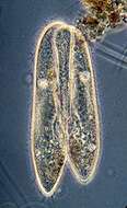
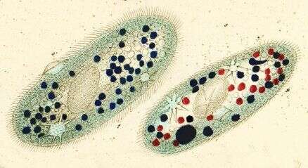
Paramecium (caudatum) (par-a-mee-see-um) is a very familiar genus of ciliates. They eat bacteria and have the mouth recessed in a buccal cavity, and the cell is often shaped with a scoop leading to the mouth. There are cilia all over the body with a caudal tuft of longer cilia at the back of the body. Usually with a layer of extrusomes (trichocysts) under the cell surface and a large oval macronucleus. Contractile vacuoles star-shaped. Most species are elongate, although this particular individual has been squashed so that we can see the nuclei. The (morpho)species are distinguished by morphology of the nuclei. This species is P. caudatum, which a single rather globular macronucleus lying alongside (4 o clock) the macronucleus. Phase contrast.
-
This cell was encountered around the margins of the alkaline Mono Lake. Paramecium is not known to occur in extreme habitats, but as the marginal regions of the pond receive run-off from the adjacent land, we may presume that it lived in a less extreme micro-habitat. It does, however, seem to be eating Picocystis - a common picophytoplankton organism in Mono Lake. Some extrusomes have been expelled from the central region of the cell. Phase contrast micrograph.
-
Paramecium (caudatum) (par-a-mee-see-um) is a very familiar genus of ciliates. They eat bacteria and have the mouth recessed in a buccal cavity, and the cell is often shaped with a scoop leading to the mouth. This cell has been compressed so that we can see various organelles. There are cilia all over the body with a caudal tuft of longer cilia at the back of the body. Usually with a layer of extrusomes (trichocysts) under the cell surface and a large oval macronucleus. Contractile vacuoles star-shaped. Phase contrast.
-
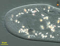
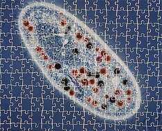
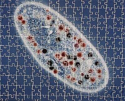
This cell has been fed on stained bacteria and indian ink to monitor the rate of food vacuole formation. Oh, and we made a jigsaw puzzle out of the picture.
-
-
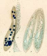
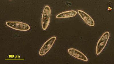
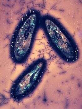
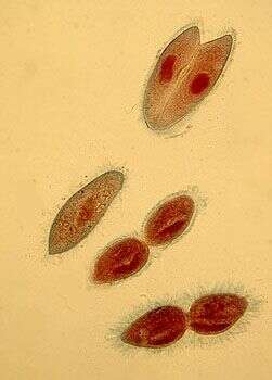
These three individuals of this small species of Paramecium have been allowed to dry in a suspension of nigrosin. The stain dries around the cell, showing off the dimpling of the surface, and the ingestion area (in the lower cell we can see the arc-shaped buccal cavity. Crystals inside the cell appear light blue. Dark lines outside the cell are trichocysts (a type of extrusome) that have been expelled from the cells.
-

Detail of the oral apparatus and surrounding infraciliature (anterior ventral aspect) of the hymenostome ciliate Paramecium caudatum (Ehrenberg, 1833). The preoral and postoral sutures (lines of convergence of kinetal fields) can be clearly seen. The kinetids with their kinetodesmal fibers are clearly visible. The kinetodesmal fibers are periodically striated bundles of fibrils arising near the base of somatic kinetids (the posterior one if kinetids are paired) in ciliates. The kinetodesmal fibers extend anteriorly and to the right of their kinety (this is the Law of desmodexy). This provides a means of determining an anterior, posterior and right/left orientation in ciliates. Here the kinetodesmal fibers are longer than the interkinetal distance and therefore overlap giving the appearance of a longitudinal interkinetal line. The macronucleus and the single large micronucleus are seen anterior to the oral aperture. Silver carbonate stain (see Foissner, W.Europ. J. Protistol.27,313-330;1991). Specimen collected from freshwater pond near Boise, Idaho July 2004. Brightfield.
-
-
These two individuals have fused toigether and wille xchange haploid nuclei as part of a process of sexual activity. Ciloiates can divide asexually with mitotic division of the micronuclei and an a-mitoitic division of the macronucleus. Periodically, the cells go through a sexual process in which cells join at the mouth region, the macronucleus breaks down, micronuclei undergo meiosis, haploid nuclei are exchanged, they then fuse and the resulting product divides to create new micronuclei and macronuclei.
-
-
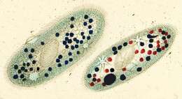
In this stained preparation, the nuclei are red. There is a single normal cell, a pair of cells joined in conjugation and two cells in the process of asexual division - which involves a division furrow cutting through the middle of the cells.
-
-
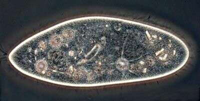
This single cell has been fixed and stained with a dye that shows up the nuclei. The image clearly shows the large macronucleus and the smaller, but still substantial. micronucleus. The arrangement of the nuclei is one of the criteria used to distinguish the species in this genus.
-
-
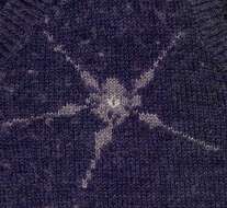
Passion. This image goes with the one of Eufolliculina. In this case this is a knitted contractile vacuole with the radiating canals, ampullae, vacuole and vacuolar pore. Knitted by June Hornby.