-
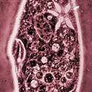
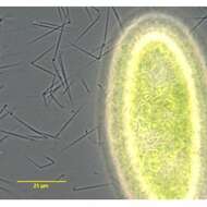
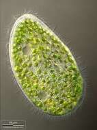
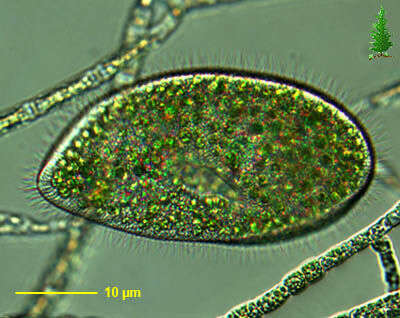
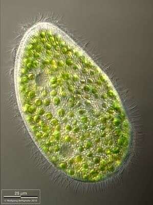
Paramecium (bursaria) (par-a-mee-see-um) is a very familiar genus of ciliates. They eat bacteria and have the mouth recessed in a buccal cavity. There are cilia all over the body with a caudal tuft of longer cilia at the back of the body. Usually with a layer of extrusomes (trichocysts) under the cell surface and a large oval macronucleus. Contractile vacuoles star-shaped. This species is P. bursaria, a species with symbiotic green algae living inside. In this image, the cell is feeding, and at rest with its cilia on the lower side inactive but holding on to the debris. Phase contrast.
-
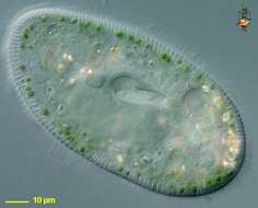
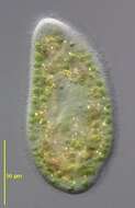
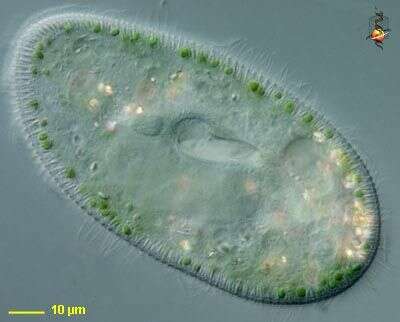
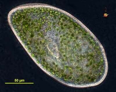
Paramecium (bursaria) (par-a-mee-see-um) is a very familiar genus of ciliates. They eat bacteria and have the mouth recessed in a buccal cavity, and the cell is often shaped with a scoop leading to the mouth. There are cilia all over the body with a caudal tuft of longer cilia at the back of the body. Usually with a layer of extrusomes (trichocysts) under the cell surface and a large oval macronucleus. Contractile vacuoles star-shaped. This species is P. bursaria, a species with symbiotic green algae living inside. Differential interference contrast.
-
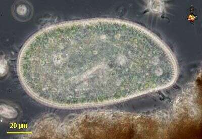
Paramecium bursaria - this species of Paramecium is squatter than the more familiar aurelia or caudatum-like species, and is also distinguished because it contains symbiotic green algae. The light structure to the left of centre is the opening to the mouth. Phase contrast micrograph.
-
Collected from Cumloden Swamp on December 23, 2003.
-

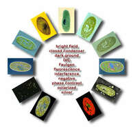
Paramecium bursaria imaged with an array of contrast enhacement techniques. All cells point towards the center of the circle. The most visible components of the cell are the large macronucleus, an ajacent micronucleus, symbiotic algae, and the mouth. Bright field (Koehler illumination - (1 o'clock) shows the algae and major organelles, and these are made more strongly visible when the condesner iris is closed (2 o'clock). Phase contast tends to generate a lot of noise inside the cell (9 o'clock), whereas Nomarski (DIC or differential interference contrast - 10 o'clock) creates an image that is like a slice through the cell. Interference optics (3 o'clock)convert elements with different optical properties into different colours. Polarising optics (8 o'clock) only show refringent crystals weithin the cell. In dark ground (7 o'clock), the object is illuminated obliquely and only refracted light is visible. Illumination with ultraviolet light causes autofluorescence of the algae and the nucleus shows up because of the addition of DAPI (5 o'clock). The nucleus is also clearly visible in the cell that has been stained with Feulgen stain (for DNA - 12 o'clock). Cells dried in indian ink have ink collected in the depressions on the cell surface and the ink collects around the cell - this is referred to as negative staining (4 o'clock). Silver stains reveal the bases of the cilia (11 o'clock). The dowload file contains intermediate sized images of each technique. Images by D . J. Patterson.
-
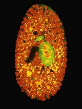
Thjis living cell has been placed in a dilute solution of DAPI stain before being observed by fluorescence microscopy. DAPI binds with DNA and looks light when illuminated with short wavelength light (ultra violet or blue) - part of the fluorescence technique. The technique involves shining low wavelength light at cells and then filtering the wavelengths out. If the light has interacted with any materials in the speciment to lead to fluorescence, this can then be seen as bright areas against the dark background. The chlorophyll in the symbiotic algae fluoresce red, while the macronucleus and micronucleus fluoresce a light green colour.
-
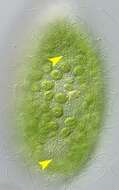
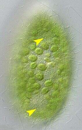
Dorsal view of Paqramecium bursaria (EHRENBERG,1831) FOCKE, 1836.The yellow arrowheads mark the single excretory pores of the two contractile vacuoles. DIC.
-
Paqramecium bursaria (EHRENBERG,1831) FOCKE, 1836.many exploded extrusomes are visible. Phase contrast.
-
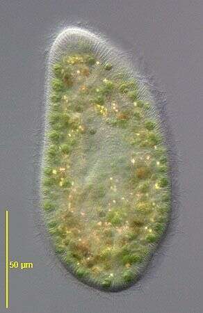
in vivo portrait of Paramecium bursaria (EHRENBERG,1831) FOCKE, 1836. DIC.
-
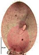
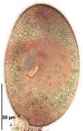
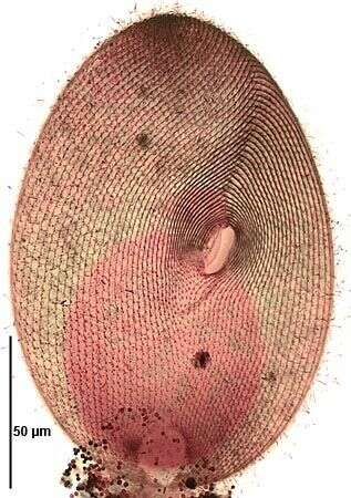
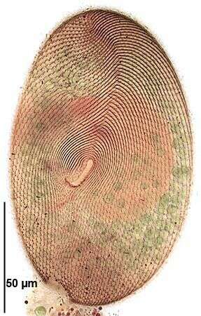
Ventral infraciliature of Paramecium bursaria (EHRENBERG,1831) FOCKE, 1836. Stained by the silver carbonate technique (Foissner,W. Europ. J. Protistol.27:313-330;1991).Brightfield.
-
Ventral infraciliature of Paramecium bursaria (EHRENBERG,1831) FOCKE,1836. Stained by the silver carbonate technique (Foissner,W. Europ. J. Protistol.27:313-330;1991).Brightfield.
-
Dorsal view of Paramecium bursaria. On top left the metachronous beating of cilia is visible. Sample from sphagnum pond situated in the northern alpine region of Austria near Salzburg. Images were taken using Zeiss Universal with Olympus C7070 CCD camera.
-
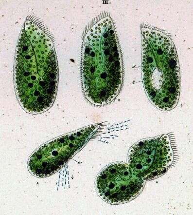
Originally described by Ehrenberg under the name Loxodes bursaria.