Translation of Schultze's original description
provided by EOL authors
Prorhynchus stagnalis nov. gen. nov. spec.
Tablet 6, figure 1.
Body 1½ ''' - 2 ''' long, 1/6 ''' wide, when expanded, cylindrical, fore-end smaller than rear-end, white. Eyes missing, the proboscis is short, not eversible but rather protrusile; therefore the weapon itself lies immediately behind the frontal opening. Residence in fresh water.
Among all the nemerteans so far known there are none whose proboscis lies so near to the fore-end of the animal, nor any whose conformation [of the proboscis] may be compared with that of this animal. For this reason I was compelled to create a new genus.
The central nervous system of nemerteans consists, as was noted by Quatrefages, Frey, and Leuckart, of two ganglia, linked to one another by twin bridges. Such a conformation have I also recognized in Tetrastemma obscurum (figure 2aa). The nervous system of Prorhynchus stagnalis also consists of two rather remarkably pale ganglia (figure 1aa), between which only one commissure was seen.
From the ganglia a nerve goes out towards the fore-end on each side to innervate the ciliated pits. The significance of this peculiar organ, with long cilia embedded in pits of the integument, and unseen in the greater part of nemerteans examined so far, is yet unclear. The strong nerve, which in many cases returns back to follow the same [?], makes probable the hypothesis that a peculiar sensory function is to be found therein. Eyes are entirely missing. A strong nerve from each ganglion likewise goes out towards the rear, and can be followed to the very rearmost part of the body.
The proboscis consists of a foremost armed part (d), and a more muscular rear part (e). The latter encloses a canal within, which widens into a bladder at the rear end. It is probable that this holds a liquid secretion, which works as a poison during wounding [of prey] with the stylet. The opening at the foremost tip of the body serves as an outlet for the proboscis. The stylet consists of a sharp, nail-shaped spike in the middle, two accessory daggers, which are each about half so long as the spike, and whose foremost parts each lie tightly against it, with their back ends somewhat sticking out away, and a cylindrical, forward-narrowing capsule for the stylet, to be pierced by the spike. The latter ones [the sheath components] consist in their foremost end of the same substance as the spike, but towards the back end change into a membranous cavity which widens at the end, which lie upon the muscular, cross-wrinkled part of the proboscis with its terminal bladder. Entirely separated from this organ is the alimentary canal. This begins with the tube-shaped pharynx (e), which lies near the proboscis, and which can be ejected some distance out, perhaps to the very foremost opening of the animal. Following this is the gut (g), running directly to the back end of the animal, which ends by (h) into an anus [note: there is actually no anus]. This is, when filled, somewhat massive [lit. buchtig, an unknown word, but perhaps really wuchtig = weighty, massive], and allows only a little bit of remaining room for the sexual organs. In its interior there are many oil droplets whose cells contain dark nuclei, with which we are familiar from the guts of Rhabdocoels. Vascular diverticulations of extraordinary delicateness, without contractile edges and with a trembling motion in the interior, therefore probably nephridia [lit. “water-vessels”], can be recognized in the anteriormost transparent part of the animal.
The few specimens found by me have all been female. A tube-shaped ovary lies near the intestine in the posterior half of the body. The smallest part of this (k), lying near to the anus, encloses an egg germs of some mass. Towards the front these are mature, and surrounded by egg mass, and individual curvatures across the length of the organ seem to indicate a tendency towards constriction of the forward part of the ovary, exactly as with Macrostomum. Several times I saw an isolated egg (i), without a hard shell, with germinal vesicles and cored yolk cells, laying in the foremost end of the ovary. Truthfully, a sexual opening was never observed.
Prorhynchus stagnalis appears but seldom in Greifswald, in a great pond with a peaty bottom, on the way to the village of Wackerow. At the beginning of April I found young specimens bearing not yet even a hint of sexual parts. At the end of April and August were several sexually mature females observed.
The numerous so far known animals that we call nemertean species all stem from the sea. Two turbellarians described by Dugès seem, however, to be exceptions; these were found in rivers from the interior of France, as Prostoma lumbricoideum and P. clepsinoideum. The first was mentioned by Ehrenburg in the Abhandlung der Academie des Wissenschafts zu Berlin 1835 pg. 244 as Tetrastemma lumbricoideum. The bodily construction and the number of eyes of both of these species, as well as the existence of an anus, proves the hypothesis that what Dugès had observed were nemerteans. For the latter species this supposition has been fully confirmed by the observations of my friend Dr. F. Müller, which he has conveyed to me orally. He has found in Tetrastemma lumbricoideum collected in the vicinity of Berlin a proboscis with a stylet and reserve stylet sacs which is characteristic of nemerteans. We must now add to this, the only certain freshwater nemertean, a second, Prorhynchus stagnalis. F. Müller has observed a third type in a peaty ditch of a moor near Greifswald. This one was about 3 ''' long, white, and had in the interior of the body a recognizable stylet with reserve spikes. Unfortunately no drawings of the only observed specimens were ever completed. They could not again be found later.
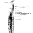
Tablet 6
1 – Prorhynchus stagnalis nov. gen. nov. spec., a freshwater nemertean. aa. Both central ganglia of the nervous system. b.b. Ciliated pits. c. Opening for the extension of the proboscis, possibly also the mouth opening. d. Forwardmost armed part of the proboscis. e. Rearmost, muscular part of the same. f. Tube-formed, muscular pharynx. g. Intestine. h. Anal opening. i. An egg with germinal vesicles and yolk cells. k. The lower part of the ovary, filled with egg germs.