-
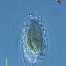
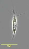
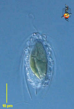
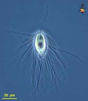
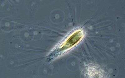
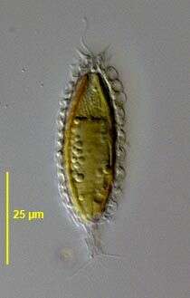
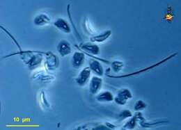
Mallomonas (ma-la-moan-ass) is a synurophyte alga, traditionally regarded as a chrysophyte or related to the chrysophytes, but distinguished by the presence of extracellular siliceous scales. This genus has large chlorophyll a and c containing plastids, an apical flagellum, and a coating of slipper shaped scales each of which has a long projecting spine. Differential interference contrast.
-
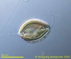
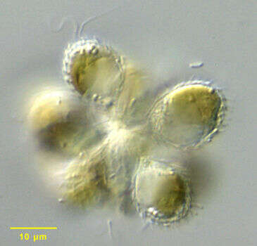
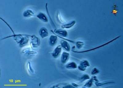
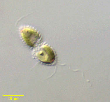
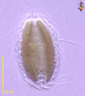
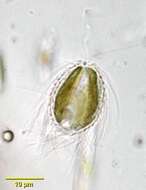
Synura sphagnicola, a colonial chrysophyte. Synonymous with Skadovskiella. Cells are joined at their posterior ends in the center of the colony. Cells are ovoid with two golden chloroplasts. There are two equal flagella less than 1 cell length. Siliceous scales with a unique ring form from which there is a rod-like projection cover the cell. A detached scale is seen in the center of this image. The scale structure is diagnostic for the species. A stigma is absent. Large vacuoles containing the glucopyranoside storage polymer, chrysolaminarin (leucosin) accumulate in the cytoplasm . The colonies swim with a slow rolling motion.From a polysaprobic temporary freshwater farm pond near Boise, Idaho. Differential interference contrast.Differential interference contrast optics.
-
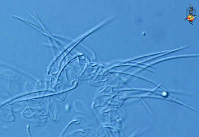
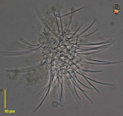
Mallomonas (ma-la-moan-ass) is a synurophyte alga, traditionally regarded as a chrysophyte or related to the chrysophytes, but distinguished by the presence of extracellular siliceous scales. This genus has large chlorophyll a and c containing plastids, an apical flagellum, and a coating of slipper shaped scales each of which has a long projecting spine. This image is a detail of the empty periplast (coating of scales and spines) and showing the two kinds of scales which make up the periplast. Differential interference contrast.
-
Synura sphagnicola, a colonial chrysophyte. Synonymous with Skadovskiella. Cells are joined at their posterior ends in the center of the colony. Cells are ovoid with two golden chloroplasts. There are two equal flagella less than 1 cell length. The cell is covered by siliceous scales the morphology of which is diagnostic for the species (see accompanying image). A stigma is absent. Large vacuoles containing the glucopyranoside storage polymer, chrysolaminarin (leucosin) accumulate in the cytoplasm. The colonies swim with a slow rolling motion.From a polysaprobic temporary freshwater farm pond near Boise, Idaho. Differential interference contrast.
-
Mallomonas (ma-la-moan-ass) is a synurophyte alga, traditionally regarded as a chrysophyte or related to the chrysophytes, but distinguished by the presence of extracellular siliceous scales. This genus has large chlorophyll a and c containing plastids, an apical flagellum, and a coating of slipper shaped scales each of which has a long projecting spine. Differential interference contrast.
-
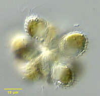
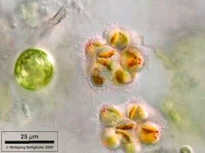
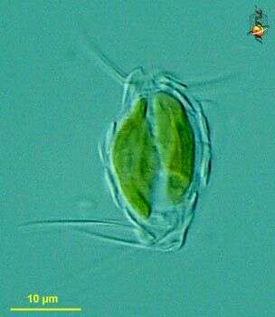
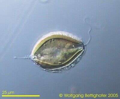
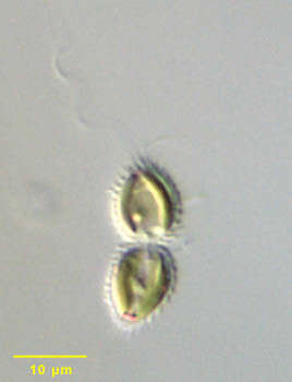
Synura sphagnicola can be preferentially found in sphagnum ponds. Typical are the reddish oil droplets they bear in plasma. Sample from sphagnum pond Dosenmoor near Neumuenster (Schleswig-Holstein, Germany). This image was taken using Zeiss Universal with Olympus C7070 CCD camera.
-
Mallomonas (ma-la-moan-ass) is a synurophyte alga, traditionally regarded as a chrysophyte or related to the chrysophytes, but distinguished by the presence of extracellular siliceous scales. This genus has large chlorophyll a and c containing plastids, an apical flagellum, and a coating of slipper shaped scales each of which has a long projecting spine. This image is of the empty periplast (coating of scales and spines). Phase contrast.
-
Mallomonas (ma-la-moan-ass) is a synurophyte alga, traditionally regarded as a chrysophyte or related to the chrysophytes, but distinguished by the presence of extracellular siliceous scales. This genus has large chlorophyll a and c containing plastids, an apical flagellum, and a coating of slipper shaped scales each of which has a long projecting spine. Phase contrast.
-
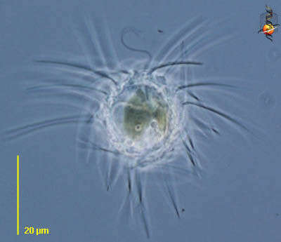
Mallomonas (ma-la-moan-ass) is a synurophyte alga, traditionally regarded as a chrysophyte or related to the chrysophytes, but distinguished by the presence of extracellular siliceous scales. This genus has large chlorophyll a and c containing plastids, an apical flagellum, and a coating of slipper shaped scales each of which has a long projecting spine. Differential interference contrast.
-
Mallomonas (ma-la-moan-ass) is a synurophyte alga, traditionally regarded as a chrysophyte or related to the chrysophytes, but distinguished by the presence of extracellular siliceous scales. This genus has large chlorophyll a and c containing plastids, an apical flagellum, and a coating of slipper shaped scales each of which has a long projecting spine. Phase contrast.
-
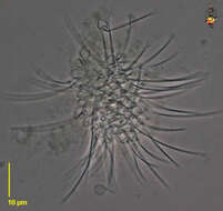
Mallomonas (ma-la-moan-ass) is a synurophyte alga, traditionally regarded as a chrysophyte or related to the chrysophytes, but distinguished by the presence of extracellular siliceous scales. This genus has large chlorophyll a and c containing plastids, an apical flagellum, and a coating of slipper shaped scales each of which has a long projecting spine. The spines are particular well developed in this species. Differential interference contrast.
-
Mallomonas (ma-la-moan-ass) is a synurophyte alga, traditionally regarded as a chrysophyte or related to the chrysophytes, but distinguished by the presence of extracellular siliceous scales. This genus has large chlorophyll a and c containing plastids, an apical flagellum, and a coating of slipper shaped scales each of which has a long projecting spine. The spines are particular well developed in this species. Phase contrast.
-
Mallomonas (ma-la-moan-ass) is a synurophyte alga, traditionally regarded as a chrysophyte or related to the chrysophytes, but distinguished by the presence of extracellular siliceous scales. This genus has large chlorophyll a and c containing plastids, an apical flagellum, and a coating of slipper shaped scales each of which has a long projecting spine. This image is of a number of those scale-spine combinations. Phase contrast.
-
Mallomonas (mall-owe-moan-ass) a stramenopile with golden plastids, and with scales and spines on the outside of the cell. Differential interference microscopy.
-
-
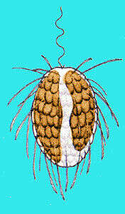
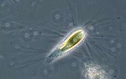
Mallomonas, a flagellate included in the Synurophyceae. Mallomonas is a very large genus with over 120 recognized species. Cells are solitary and biflagellate although the short flagellum is usually not visible by light microscopy. Chloroplasts are either double or single and bilobed. The chloroplasts of Mallomonas differ from those of chrysophytes in that they contain chlorophyll a and c1 but not chlorophyll c2. A red stigma is absent. There is a posterior contractile vacuole. Cells are covered by siliceous scales and bristles. Both are formed intracellularly then extruded to the cell surface where the bristles then articulate with or adhere to the scales. Taxonomy is based on scale and bristle ultrastructure. There are some species which form siliceous cysts known as stomatocysts. These have a terminal opening or porus which is sealed with a plug. From a freshwater pond near Boise, Idaho. Phase contrast.
-
Mallomonas, a very large genus with over 120 recognized species. Cells are solitary and biflagellate although the short flagellum is usually not visible by light microscopy. Chloroplasts are either double or single and bilobed. The chloroplasts of Mallomonas differ from those of chrysophytes in that they contain chlorophyll a and c1 but not chlorophyll c2. A red stigma is absent. There is a posterior contractile vacuole. Cells are covered by siliceous scales and bristles. Both are formed intracellularly then extruded to the cell surface where the bristles then articulate with or adhere to the scales. Taxonomy is based on scale and bristle ultrastructure. There are some species which form siliceous cysts known as stomatocysts. These have a terminal opening or "porus" which is sealed with a plug. From a freshwater pond near Boise, Idaho. Brightfield illumination
-
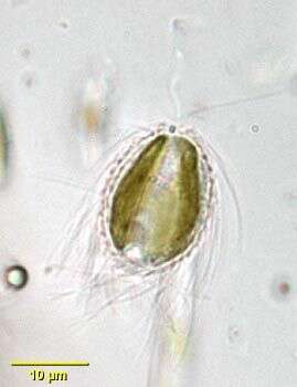
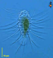
Monadic species of Synurophyceae. Cell is covered by siliceous scales. Further pictures showing also trailing flagellum in ZIP archive. Collected from littoral region (stand of Phragmites) of a rain storage reservoir in Kiel (Schleswig-Holstein, Germany). Images were taken using Zeiss Universal with Olympus C7070 CCD camera.
-
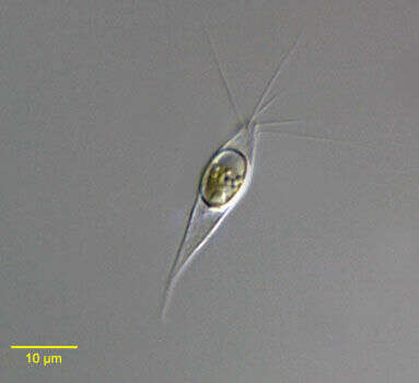
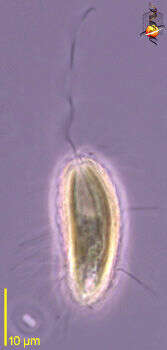
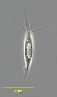
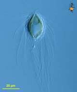
Portrait of an encysted individual of the synurophyte flagellate Mallomonas akrokomos (Ruttner in Pascher,1913). Mallomonas is a very large genus with over 120 recognized species. Cells are solitary and biflagellate although the short flagellum is usually not visible by light microscopy (no flagellum is seen in this encysted cell). Chloroplasts are either double or single and bilobed. The chloroplasts of Mallomonas differ from those of chrysophytes in that they contain chlorophyll a and c1 but not chlorophyll c2. A red stigma is absent. Siliceous scales and bristles cover cells. Both are formed intracellularly then extruded to the cell surface where the bristles then articulate with or adhere to the scales. Taxonomy is based on scale and bristle ultrastructure. In M. akrokomos, only a few anterior apical siliceous scales bear long serrated bristles almost the length of the cell. Small flat scales cover the body (not seen in this image). From a freshwater pond near Boise, Idaho. DIC optics.
-
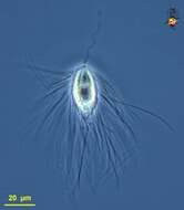
Portrait of the synurophyte flagellate Mallomonas akrokomos (Ruttner in Pascher,1913). Mallomonas is a very large genus with over 120 recognized species. Cells are solitary and biflagellate although the short flagellum is usually not visible by light microscopy. Chloroplasts are either double or single and bilobed. The chloroplasts of Mallomonas differ from those of chrysophytes in that they contain chlorophyll a and c1 but not chlorophyll c2. A red stigma is absent. Siliceous scales and bristles cover cells. Both are formed intracellularly then extruded to the cell surface where the bristles then articulate with or adhere to the scales. Taxonomy is based on scale and bristle ultrastructure. In M. akrokomos, only a few anterior apical siliceous scales bear long serrated bristles almost the length of the cell. Small flat scales cover the body. From a freshwater pond near Boise, Idaho. Phase contrast.
-
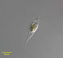
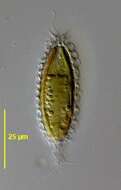
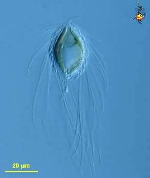
Mallomonas insignis (Penard, 1919). M. insignis has ovoid siliceous scales. Scales arranged around the flagellar opening bear short, sharp spines. Scales at the posterior end of the tail bear long fine spines. M. insignis is similar to M. mesolepis (Skuja) which lacks terminal spinous projections and has diamond-shaped scales. Collected from a temporary slow-flowing freshwater stream near Boise, Idaho. May 2005. DIC.
-
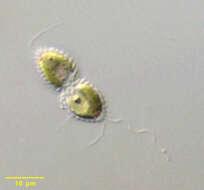
Portrait of Chrysodidymus synuroideus, a colonial synurophyte chrysophyte. Chrysodidymus is a monospecific genus. Two cells united at their bases form the colonies. The cells are conical. Each is covered by siliceous scales. The scales are formed in the chloroplast endoplasmic reticulum and transported to the cell surface. There are two flagella, one long (bearing tripartite hairs typical of stramenopiles on electron microscopy) and one short. There are two golden-colored parietal chloroplasts without pyrenoids. A posterior storage vacuole may contain chrysolaminarin. Red pigment granules can be seen at the apices of these cells. Swimming movement is a distinctive back and forth motion. From a freshwater irrigation ditch near McCall, Idaho. Differential interference contrast.
-
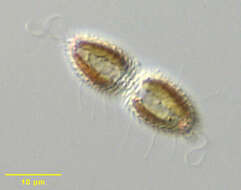
Portrait of Chrysodidymus synuroideus, a colonial synurophyte chrysophyte. Chrysodidymus is a monospecific genus. Two cells united at their bases form the colonies. The cells are conical. Each is covered by siliceous scales, some bearing spines (seen well in this image). The scales are formed in the chloroplast endoplasmic reticulum and transported to the cell surface. There are two flagella, one long (bearing tripartite hairs typical of stramenopiles on electron microscopy) and one short. There are two golden-colored parietal chloroplasts without pyrenoids. A posterior storage vacuole may contain chrysolaminarin. Red pigment granules can be seen at the apices of these cells. Swimming movement is a distinctive back and forth motion. From a freshwater irrigation ditch near McCall, Idaho. Differential interference contrast
-
Portrait of Chrysodidymus synuroideus, a colonial synurophyte chrysophyte. Chrysodidymus is a monospecific genus. Two cells united at their bases form the colonies. The cells are conical. Each is covered by siliceous scales. The scales are formed in the chloroplast endoplasmic reticulum and transported to the cell surface. There are two flagella, one long (bearing tripartite hairs typical of stramenopiles on electron microscopy) and one short. There are two golden-colored parietal chloroplasts without pyrenoids. A posterior storage vacuole may contain chrysolaminarin. Red pigment granules can be seen at the apices of these cells. Swimming movement is a distinctive back and forth motion. From a freshwater irrigation ditch near McCall, Idaho. Differential interference contrast.