-
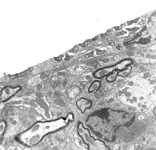
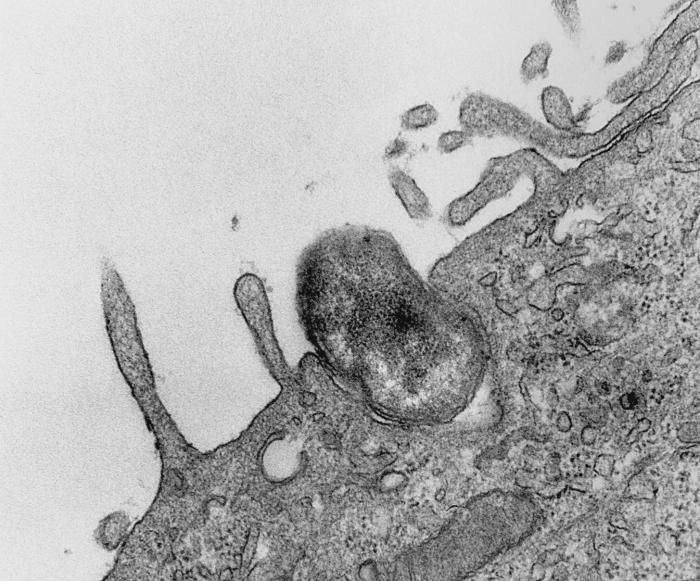
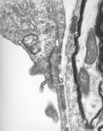
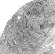
This 1976 transmission electron micrograph (TEM) depicted a hypertrophic peritoneal mesothelial cell from a mouse that had been experimentally infected intraperitoneally with Orientia tsutsugamushi rickettsial micro-organisms. This micrograph captured an organism as it appeared within a phagocytic vacuole, still bearing a third outer membrane layer of probable host cell origin.Created: 1976
-
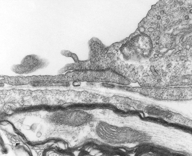
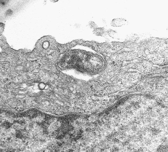
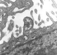
This 1976 transmission electron micrograph (TEM) depicted a peritoneal mesothelial cell from a mouse that had been experimentally infected intraperitoneally with Orientia tsutsugamushi rickettsial micro-organisms. In this particular field of view, an organism was photographically captured as it was budding from the luminal cell surface, while still covered by a third layer consisting of the host cell's plasma membrane.Created: 1976
-
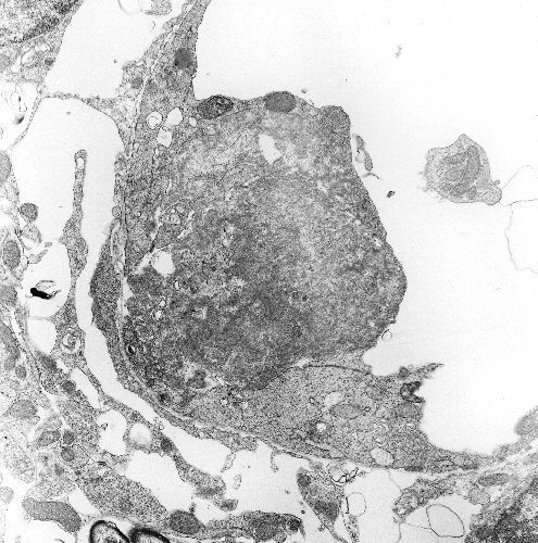
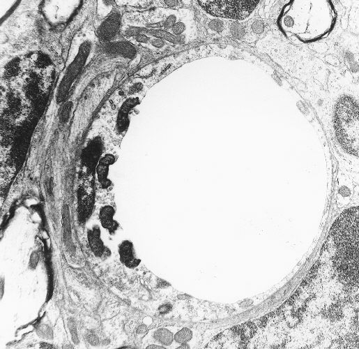
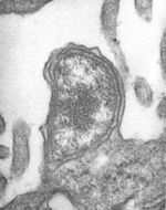
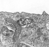
This 1978 transmission electron micrograph (TEM) depicted a brain capillary of mouse that had been experimentally infected intravenously with Orientia tsutsugamushi rickettsial micro-organisms. In this particular field of view, a hypertrophic capillary endothelial cell contained one visible organism free within its cytoplasm. The adjacent endothelial cell was of normal thickness.Created: 1978
-
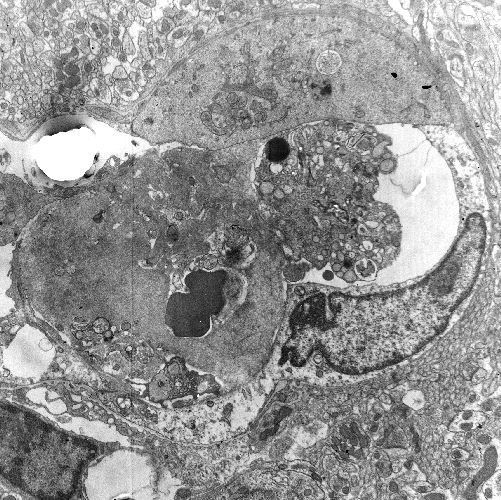
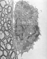
This 1976 transmission electron micrograph (TEM) depicted a brain capillary of a mouse that had been experimentally infected intravenously with Orientia tsutsugamushi rickettsial micro-organisms. Adhering to the luminal surface of the capillary's endothelium was a large macrophage containing multiple organisms free within its cytoplasm.Created: 1978
-
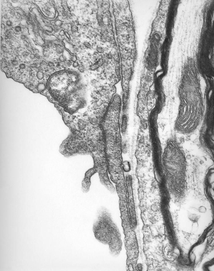
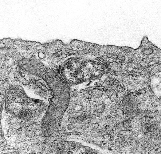
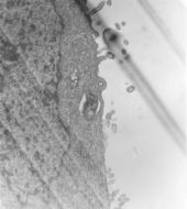
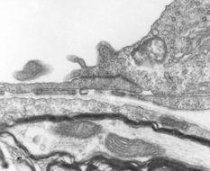
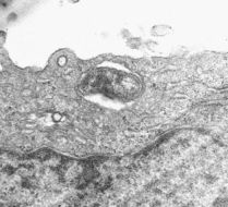
This 1976 transmission electron micrograph (TEM) depicted an extracellular Orientia tsutsugamushi rickettsial micro-organism, covered with a distinct third outer membrane of probable host mesothelial cell origin. The specimen from which this image was obtained, was extracted from the peritoneal cavity of experimentally infected mouse.Created: 1976
-
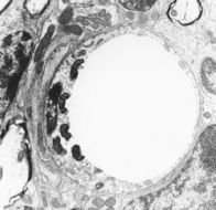
Thrombus due to infection with Rickettsia tsutsugamushi, mouse brain capillary.Created: 1977
-
Thrombus due to infection with Rickettsia tsutsugamushi, mouse brain capillary.Created: 1977
-
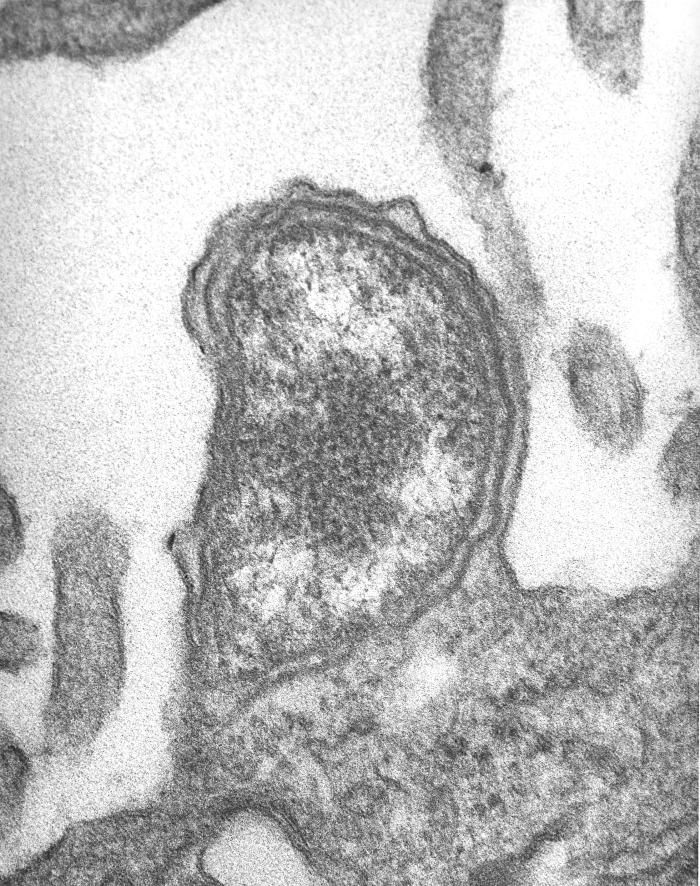
Single Rickettsia tsutsugamushi free within the cytoplasm of a mouse brain capillary endothelial cell.Created: 1977
-
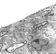
Multiple Rickettsia tsutsugamushi free within the cytoplasm of a mouse brain capillary endothelial cell.Created: 1977
-
Multiple Rickettsia tsutsugamushi free within the cytoplasm of a mouse brain capillary endothelial cell.Created: 1977
-
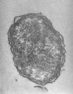
Multiple Rickettsia tsutsugamushi free within the cytoplasm of a mouse peritoneal mesothelial cell.Created: 1976
-
Rickettsia tsutsugamushi within the remains of a phagocytic vacuole of mouse peritoneal mesothelial cell.Created: 1976
-
Rickettsia tsutsugamushi within intact phagocytic vacuole of mouse peritoneal mesothelial cell.Created: 1976
-
Phagocytosis of Rickettsia tsutsugamushi by mouse peritoneal mesothelial cell.Created: 1976
-
Rickettsia tsutsugamushi budding from mouse peritoneal mesothelial cell.Created: 1976
-
Etiologic Agents of EhrlichiosesCreated: 1997
-
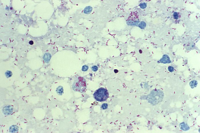
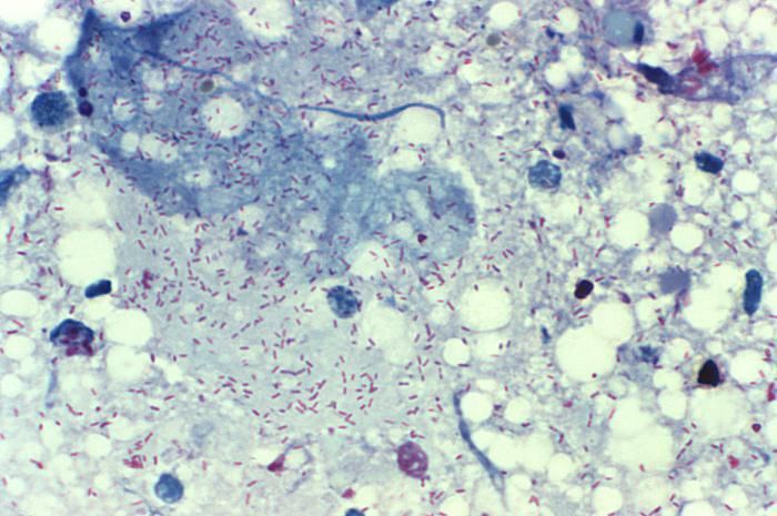
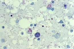
This photomicrograph of a Gimenez-stained yolk sac smear revealed the presence of Rickettsia rickettsii bacteria, which are the cause of Rocky Mountain spotted fever (RMSF). These bacteria range in size from 0.2 x 0.5 micrometers to 0.3 x 2.0 micrometers. They are difficult to see in tissues by using routine histologic stains, and generally require the use of special staining methods, such as the Gimenez stain used in this case.Created: 1974
-
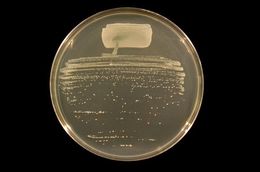
he bacterium Rhizobium tropici strain BR816 streaked to single colonies on Tryptone-Yeast Extract (TY) agar.From
Wikimedia Commons
-
-
-
-


