-
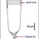
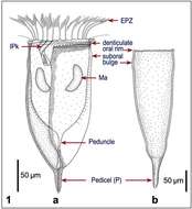
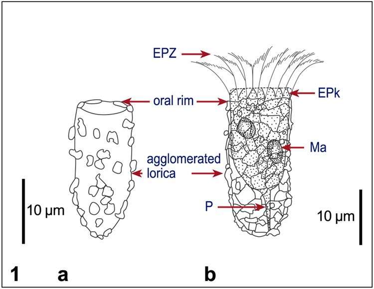
Fig 1: Tintinnopsis nana Line drawings a: Original drawing from Lohmann,1908; b. Drawing from a Lugol?s fixed specimen.
-
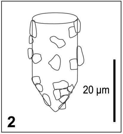
Fig 2: Tintinnopsis nana - Schematic drawing of lorica morphology, after Kofoid & Campbell, 1929
-
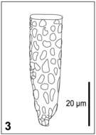
Fig 3: Tintinnopsis nana - Schematic drawing of lorica morphology, after Marshall, 1969
-
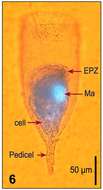
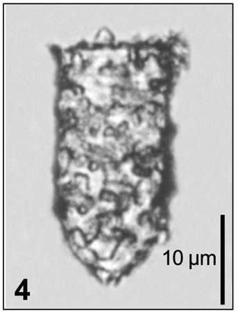
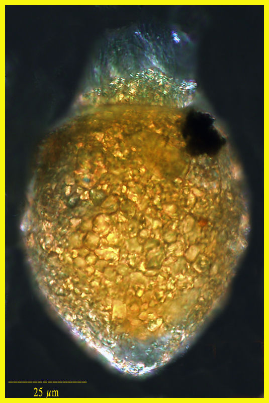
Fig 4: Tintinnopsis nana - Lugol?s fixed cell, lateral view, lorica morphology.
-
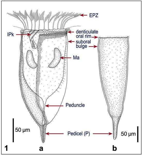
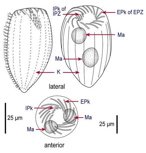
Fig 1a : Favella serrata Line drawing, showing lorica structure, and cell morphology; Fig 1b: Favella serrata Drawing of original description after Möbius (1887)
-
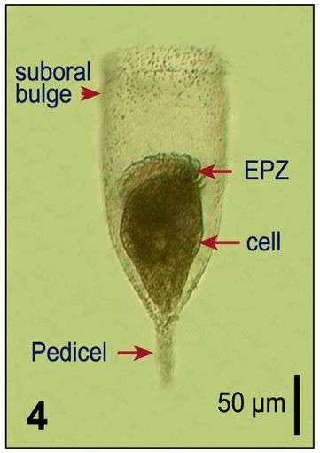
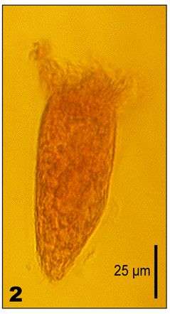
Fig 4: Favella serrata Lugol?s fixed cell, lateral view, showing pedicel, peduncle, oral rim, and ciliate cell
-
Found in a sample from the Bering Sea taken by Diane Stoecker.
-
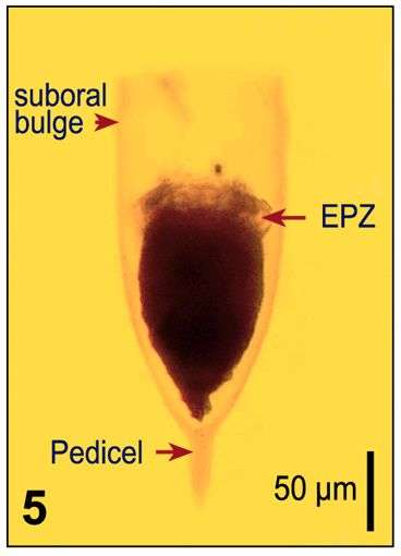
Fig 5: Favella serrata Lugol?s fixed cell, lateral view, showing pedicel, peduncle, oral rim, and ciliate cell
-
-
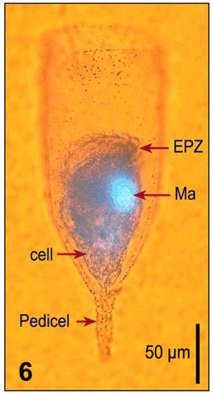
Fig 6: Favella serrata Lugol?s fixed and DAPI stained cell, illustrating shape and location of the macronuclei
-
Lugol's-fixed specimen from the Bay of Villefranche in Feb 2003
-
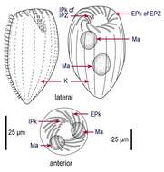
Fig 1: Strombidinopsis acuminatum Line drawing of protargol stained cells, showing kineties, oral structures and nuclei
-
Specimen found in the Bay of Villefranche in April 2010
-
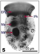
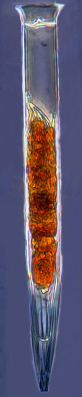
Fig 2: Strombidinopsis acuminatum Lugol's fixed cell
-
Specimen from the Etang de Thau (Sète, France) in May 2012.
-
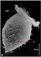
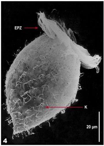
Fig 4: Strombidinopsis acuminatum SEM of Lugol's fixed cell
-
Specimen from the Etang de Thau (Sète, France) in May 2012.
-
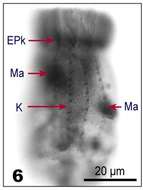
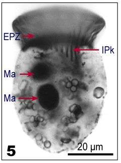
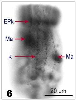
Fig 5: Strombidinopsis acuminatum protargol stained cell, lateral view: Nuclei and internal polykinetids
-
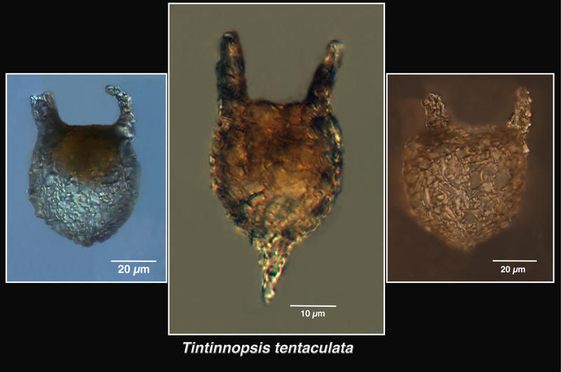
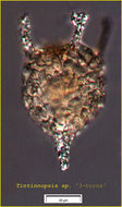
Tintinnopsis tentaculata specimens from the Ganges River estuary.
-
Fig 6: Strombidinopsis acuminatum protargol stained cell: Cell surface, showing the kineties
-
From an Indian mangrove system. All the specimens had 2 or 3 horns. The species was described by Nie & Cheng in 1947 from coastal waters of China as having 5 or 6 projections.
-
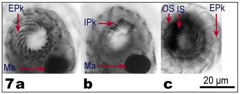
Fig 7a-c: Strombidinopsis acuminatum Apical view of protargol-stained cells, showing details of the oral structures: a. Above the cell (showing the cilia of the EPk); b. At the base of the cilia, showing the EPk; c. Within the cell (showing the IPk), OS - outer segment, IS - inner segment
-
-
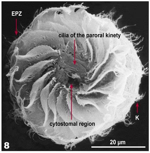
Fig 8: Strombidinopsis acuminatum SEM image, apical view of the oral region