-
El Maillo, Castille and Leon, Spain
-
Miranda do Douro Municipality, Braganca, Portugal
-
Matalebreras, Castille and Leon, Spain
-
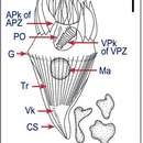
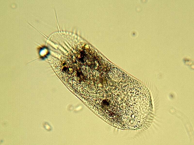
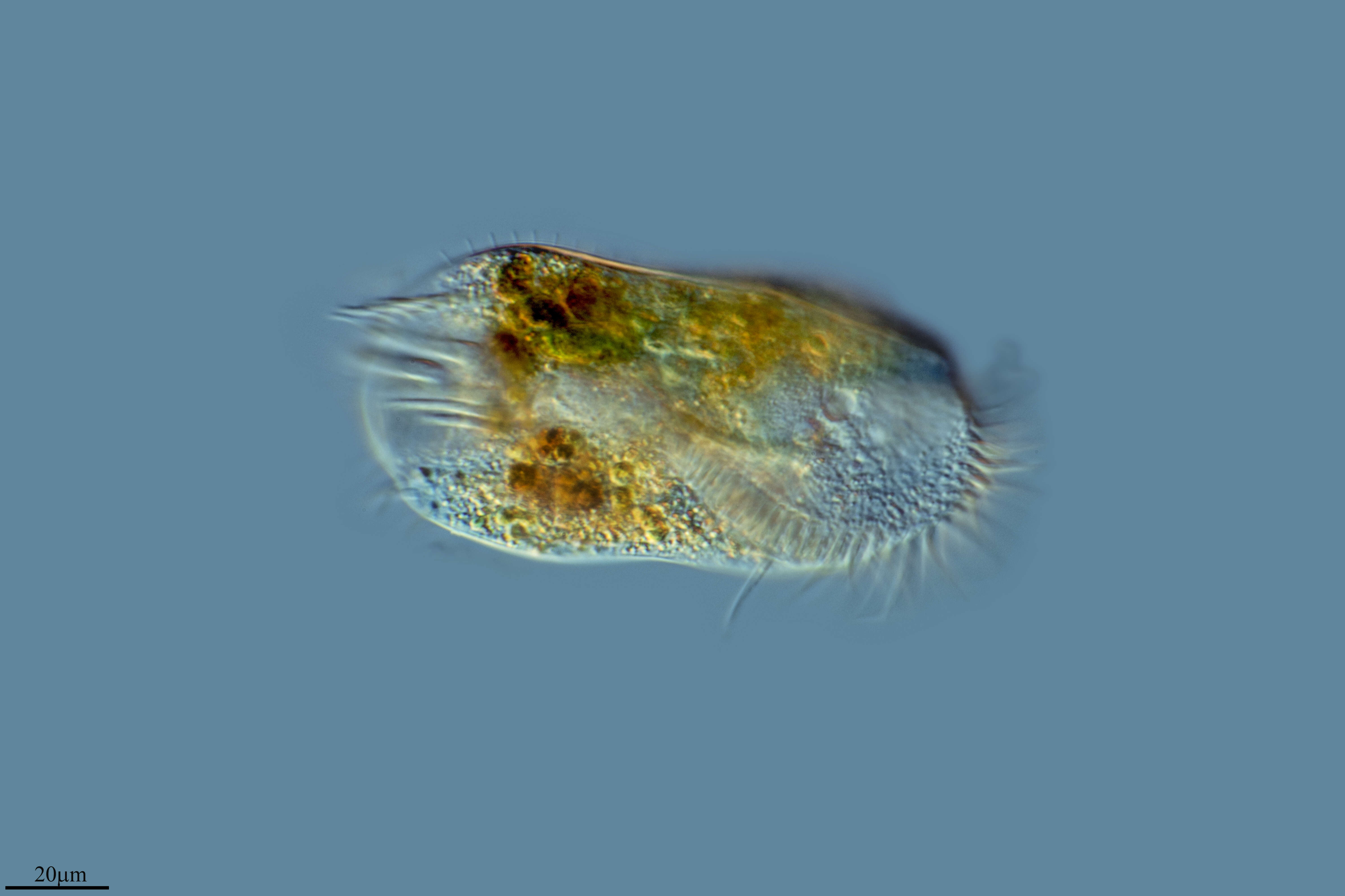
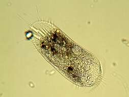
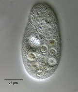
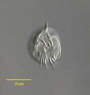
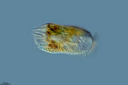
The cell body measures 100-300 micron in lenght and is dorsoventrally flattened, with a rounded anterior end and a slyghtly pointed posterior end. It has two ovoid macronuclei and two spherical micronuclei. The somatic ciliature consists of 8 frontal cirri, 5 trasverse cirri, 3 caudal cirri, 20-25 right marginal cirri and 15 left marginal cirri ( Tuffrau, 1965 ).
-
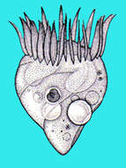
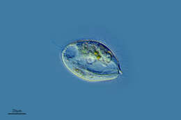
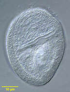
Fig 3: Lugol?s fixed cells: ventral view of normal specimens
-
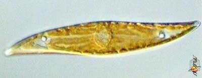
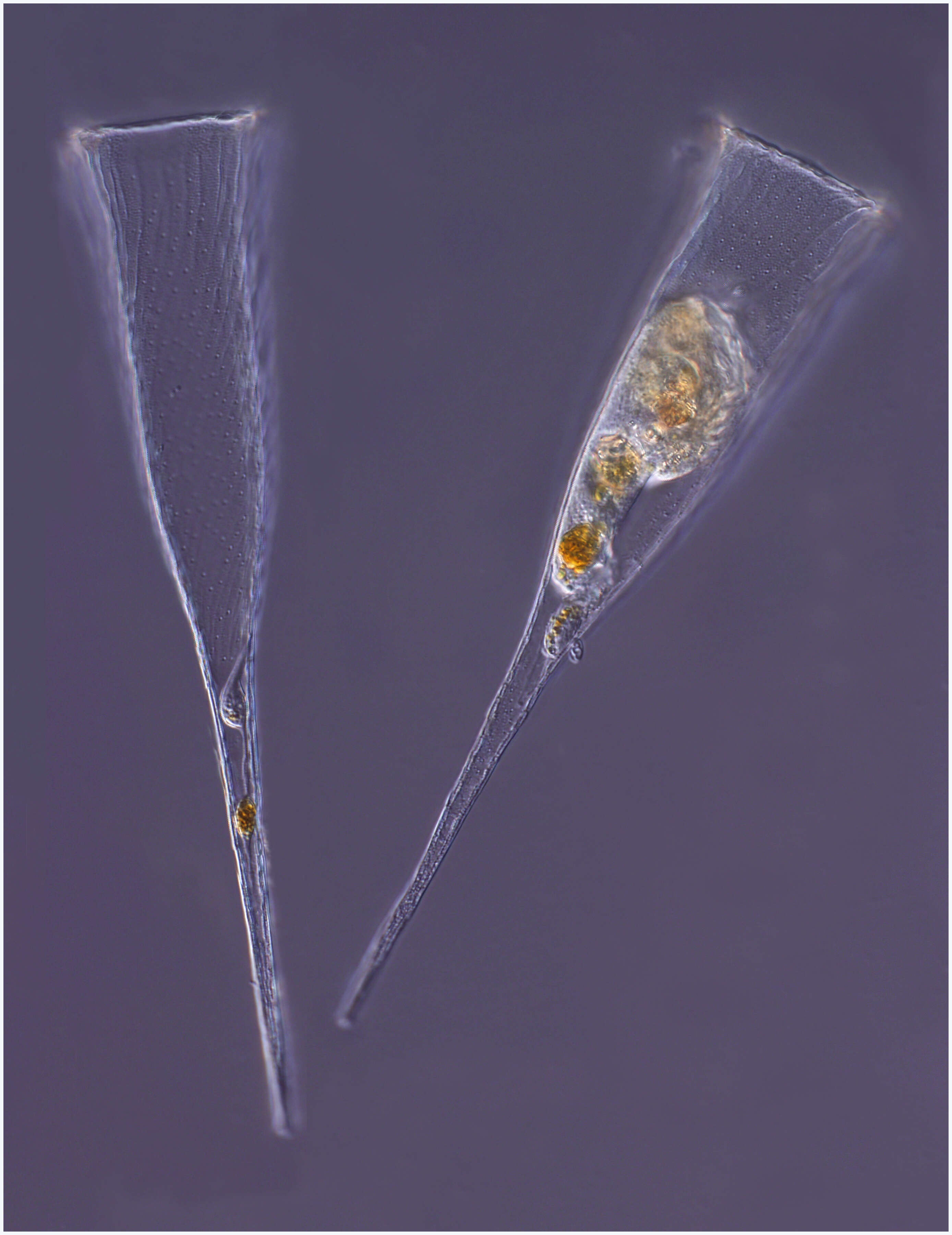
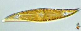
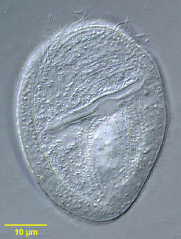
Pleurosigma (ploo-row-sig-ma) and Gyrosigma are two rather similar genera of sigmoid-shaped pennate diatoms found in intertidal sediments, salt marshes and so on. The nucleus is located at the centre of the cell. The plastids contain chlorophylls a and c which gives the yellowy-brown colour. . Pleuosigma is distinguished in part by the angled pattern of marks on the valve of the frustule. Differential interference contrast.
-
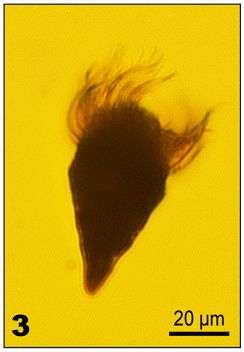
Portrait of Monodinium balbianii (FABRE-DOMERGUE,1888) in mid-division. Dividers are easily confused with Didinium species but may be distinguished by the furrow associated with the ciliary girdle of the opisthe (posterior daughter cell). Stomatogenesis is telokinetal (i.e. new oral ciliature develops from anterior ciliary rows of opisthe). Collected from organically enriched freshwater pond near Boise, Idaho in September 2003. DIC optics.
-
-
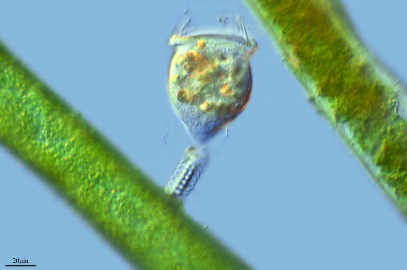
Chaetospira muelleri (LACHMAN,1856). The corkscrew shaped highly contractile anterior end of the cell is seen protriding through the opening of the flask-shaped lorica. Collected from tidal pools at Alki Beach, Seattle, Washington 47°35â41.25âN 122°23â19.60âW.January,2006. DIC.
-
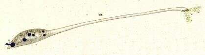
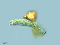
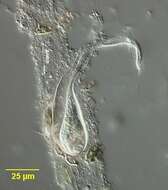
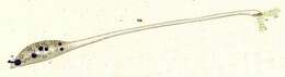
Originally described by Ehrenberg under the name Trachelocerca olor.
-
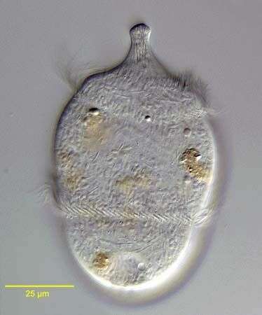
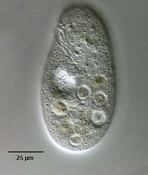
Gastronauta membranaceus (Engelmann in Bütschli,1889), a hypostome ciliate, distinguished by its long transversely oriented cytostome. The cytostome lacks trichites. The body is ovoid in outline and strongly dorsoventrally flattened. Ciliature is restricted to the ventral surface except for two short dorsal kineties anteriorly. A few somatic kineties run uninterrupted to the right of the cytostome arching around the anterior of the cell. Several right somatic kineties are interrupted by the cytostome. The left somatic kineties terminate at the cytostome. A single kinety runs around the circumference of the cytostome. An unciliated bare are overlies the region of the macronucleus posterior to the cytostome. The macronucleus is oblong and heteromerous (i.e. containing areas with markedly differing RNA and DNA contents resulting in irregular staining and optical characteristics). The single micronucleus is quite prominent. Two contractile vacuoles are present, one in the anterior half and one posteriorly. Gastronauta feeds mainly on diatoms. From a freshwater pond near Boise, Idaho. Phase contrast illumination.
-
Portrait (left anterolateral view) of the hymenostome ciliate Colpidium kleini (Foissner, 1969). Very similar in overall appearance to C. colpoda although usually more slender and with fewer somatic kineties. The cytostome is in the anterior 1/4 of the cell. There is a curved paraoral membrane along the convex right margin of the cytostome. The left margin is slightly concave. There are three adoral membranelles. There are 32 to 44 somatic kineties. The kineties to the right and left of the oral aperture meet at a curved preoral suture. There is an anterior apical area bare of cilia. There are rows of inconspicuous mucocysts between the somatic kineties. The ellipsoid macronucleus and adjacent micronucleus are centrally located. The single contractile vacuole is located in the midbody with a single excretory pore on the right surface. The feature most clearly distinguishing Colpidium kleini from C. coploda is the silverline system (as demonstrated by silver nitrate staining). Collected from an organically enriched freshwater pond near Boise, Idaho. DIC.
-
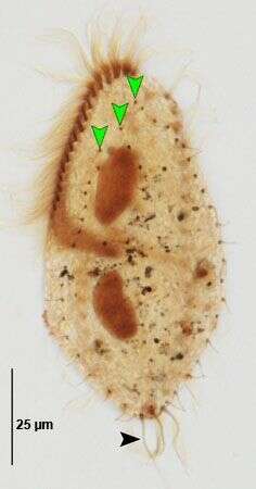
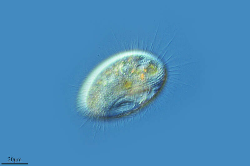
Gonostomum affine (Stein, 1859) Sterki, 1878. Dorsal kineties (green arrowheads) and caudal cirri (black arrowhead). Non-flooded Petri dish soil sample collected from flood-irrigated lawn in Boise, Idaho, June 2008. Protargol (Wilbert modification). Brightfield.
-
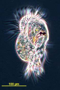
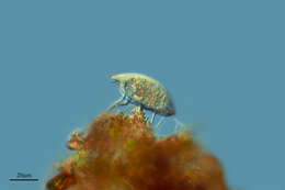
The hypotrich ciliate Uronychia isolated from Little Sippewisset Pond, Woods Hole, MA, USA. Image by Andrew Schurko.
-
Ventral view of the hypotrich ciliate, Aspidisca turrita (Ehrenberg, 1831) Claparède & Lachmann, 1858. The cytostome with adoral zone of membranelles is senn at viewer's lower right.The ventral ciiri are seen anteriorly and the transverse cirri posteriorly.Collected from a freshwater pond near Boise, Idaho. June 2005.DIC
-
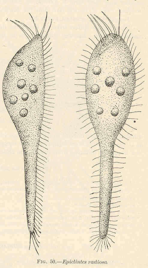
Epiclintes radiosa.
-
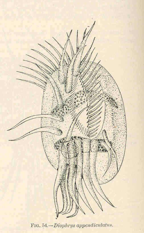
Diophrys appendiculatus.
-
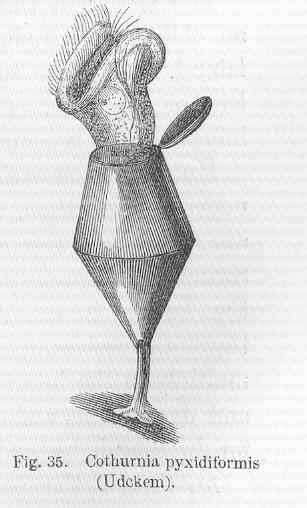
Cothurnia pyxidiformia (Udckem).
-
-
Lardero, La Rioja, Spain
-
Rancho de la Herradura, Andalusia, Spain
-
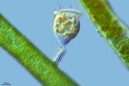
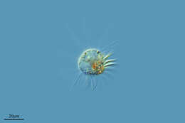
A living specimen retracted into its lorica (shell) and an empty lorica. From the Bay of Villefranche. Z-stack of photomicrographs made using a 20x objective and DIC optics.
-
Ribadelago de Franco, Castille and Leon, Spain
-
Moreruela De Tabara, Castille and Leon, Spain