-
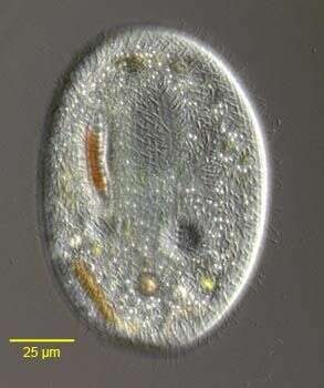
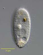
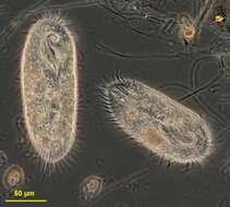
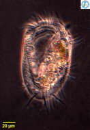
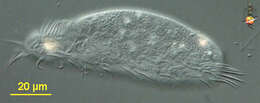
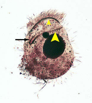
Enchelys (ench-el-is) -cylindrical predatory ciliate, body fairly flexible, mouth slit zone at anterior end, underlain by a number of extrusomes. The cell has just eaten a dinoflagellate. Differential interference contrast.
-
Chlamydodon (clam-ee-doe-don), alga-eating hypostome like many other hypostome ciliates, eats filamentous bacteria - such as filamentous blue green algae. They make contact with the filament, move along up and down until they find an end. They then tip over, pushing the end of the filament into the mouth - a cylindrical structure supported by a palisade of microtubular rods . They then start to suck the filament into the cell. As it hits the posterior margin, the cell is deformed by the stiff filament. The food is stunningly quickly degraded and begins to break and fold so that the cell can pull in a filament very much longer than itself. Yum. Phase contrast.
-

Ventral view of the infraciliature of Metopus palaeformis (Kahl,1927) contracted by fixation and compressed to display details.Synonyms probably include Tesnospira alba (Jankowski,1964),M. hyalinus (Kahl,19270 and M. tenuis (Kahl,1927) among others.Morphology is highly variable probably explaining the large number of synonyms. The cell is flask-shaped (as in this example)to elongate .The anterior end is twisted to the left resulting in a rounded lip that overhangs the peristome.The spiral peristome is bordered on the left by an adoral zone of membranelles (arrow) and on the right by five closely spaced kineties,the "perizonal stripe" (large arrowhead).Just to the right of the posterior termination of the AZM is a short, inconspicuous undulating membrane(small arrowhead).The cytoplasm contains endosymbiotic methanogenic bacilli (not seen here).Collected from the bottom sediments of an organically enriched rain pool with abundant decaying grass contaminated by Canada goose (Branta canadensis) droppings.Boise, Idaho. January 2006.Stained by the silver carbonate technique (see Foissner, W. Europ. J. Protistol., 27:313-330;1991).Brightfield.
-
-
Aspidisca, small atypical hypotrich, seen here from ventral surface. With a few ventral cirri and adoral zone of membranelles, on the right, reduced to a small brush. Differential interference contrast.
-
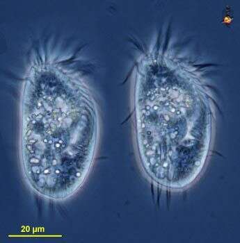
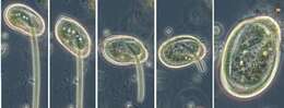
Euplotes (you-ploe-tees) is a hypotrich ciliate. There is an adoral zone of membranelles leading from the front of the cell to the mouth (where food vacuoles are formed) on the ventral side of the body. Membranelles are paddles formed by clusters of cilia which adhere to each other (at this size, water is viscous and acts like a glue holding cilia together). Hypotrichs use clumps of cilia called cirri to move. The two images (of the same cell) are taken at slightly different focal levels. Phase contrast microscopy.
-
-
-
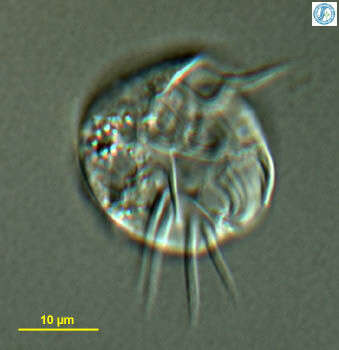
Portrait of the planktonic protstomatid ciliate, Apsiktrata gracilis (Penard,1922)Foissner, Berger & Kohmann 1994. Morphologically quite similar to members of the genus Holophrya but lacking a "dorsal brush". The anterior apical cytostome and its circumoral dikinetids is seen here.The microfibrillar system associted with the basal bodies of the somatic kineties is visible here. Collected from a freshwater pond near Boise, Idaho. Silver carbonate stain (see Foissner, W. Europ. J. Protistol., 27:313-330;1991). DIC.
-
-
Histriculus (his-trick-you-lus) is a hypotrich ciliate with cirri forming a row all around the margin and including across the back of the cell. Cell not flexible, by which it can be distinguished from the very similar Oxytricha, also not with three long caudal cirri, by which it is distinguished from Stylonychia. With adoral zone of membranelles. Phase contrast.
-
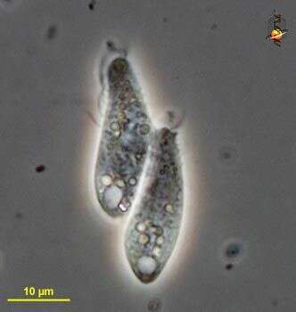
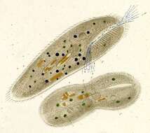
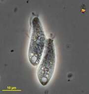
Enchelys (en-chill-iss) small predatory ciliate, here two cells are linked in conjugation (during which time DNA is exchanged - it+s a kind of sexual activity but without reproduction). Clear vacuoles at the rear are contractile vacuoles. Phase contrast micrograph.
-
-
Ventral view of the infraciliature of Metopus palaeformis (Kahl, 1927) contracted by fixation and compressed to display details...Synonyms probably include Tesnospira alba (Jankowski,1964),M. hyalinus (Kahl,19270 and M. tenuis (Kahl,1927) among others.Morphology is highly variable probably explaining the large number of synonyms. The cell is flask-shaped (as in this example) to elongate .The anterior end is twisted to the left resulting in a rounded lip that overhangs the peristome.The spiral peristome is bordered on the left by an adoral zone of membranelles (large arrowhead) and on the right by five closely spaced kineties,the "perizonal stripe" (small arrowhead).Just to the right of the posterior termination of the AZM is a short, inconspicuous undulating membrane(black arrow).The cytoplasm contains endosymbiotic methanogenic bacilli (not seen here).Collected from the bottom sediments of an organically enriched rain pool with abundant decaying grass contaminated by Canada goose (Branta canadensis) droppings.Boise, Idaho. January 2006.Stained by the silver carbonate technique (see Foissner, W. Europ. J. Protistol., 27:313-330;1991).Brightfield.
-
Thuricola (thurr-ick-owe-la) is a peritrich ciliate which lives within a lorica. Contractile and this cell has withdrawn into the lorica. A flap has closed over the contractile cell and this features distinguishes this genus. Differential interference contrast.
-
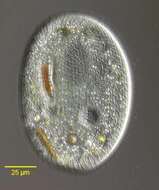
Aspidisca, a common, small hypotrich ciliate genus with many species. The cell body is rigid, colorless and dorsoventrally flattened sometimes with peripheral or dorsal spinous projections. The dorsum may be longitudinally ribbed (e.g. A. cicada). Marginal and caudal cirri are absent and ventral cirri prominent (frontoventral and transverse groups). The oral aperture is faintly visible on the organism's left posterior margin in this image (we are looking at it from the ventral side). The adoral zone of membranelles is divided into a small part at the anterior left side and a larger part around the peristome, neither is well seen in this image. Macronucleus is bipartite in some species but more usually "C" or horseshoe shaped. Usually small but one species, A. magna may exceed 150 microns in length. Probably polyphagous but mainly feeds on bacteria. From standing freshwater near Boise, Idaho. Phase contrast.
-
Euplotes, common hypotrich ciliate. There is an adoral zone of membranelles extending around the front of the cell and this is used to acquire bacteria and small protists as food. Euplotes uses cirri - collections of cilia - to walk over the substrate. From Lake Donghu, China. Phase contrast micrograph.
-
-
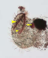
Detail of the oral aperture and ventral proboscis of Monilicaryon monilatus Monilicaryon monilatus (Stokes, 1886) Jankowski, 1967. Similar in overall appearance to Dileptus anser. M. monilatus differs by having a shorter proboscis relative to the length of the body (1/3 to 1/4) and by lacking the row of obliquely oriented closely spaced kinetids on the ventral aspect of the left side of the proboscis (this feature requires demonstration by DIC or protargol staining). M. monilatus has two single files of kinetids extending from either side of the oral aperture anteriorly along the ventral aspect of the proboscis separated by a strip bearing extrusomes (See Foissner W., Berger H and Kohmann F. Taxonomische und ökologische Revision der Ciliaten des Saprobiensystems- Band IV: Gymnostomatea, Loxodes, Suctoria. Informationsberichte Bayer. Landesamtes für Wasserwirtschaft. 1/95:185-202, 1995). In this image the two parallel kineties along the right side of the extrusome strip are visible. Collected from a freshwater pond near Boise, Idaho. DIC.
-
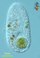
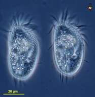
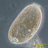
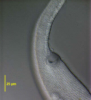
Right dorsolateral surface view of the hymenostome ciliate, Frontonia angusta (Kahl, 1931). Very similar in overall apppearance to F. acuminata (Ehrenberg,1833)Buetschli,1889. F. angusta lacks the anterior apical collection of pigmented granules seen in F. acuminata and its contractile vacuole has 3-4 excretory pores (4 in this case).The approximately 6 µm long extrusomes are clearly visible. Ingested diatoms and green algae are present. Collected from a freshwater pond near Boise, Idaho.DIC.
-
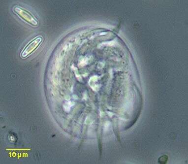
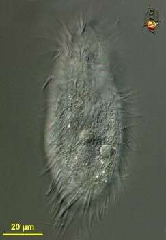
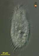
Holosticha (whole-o-stike-a) (tentative identification) is a hypotrich ciliate in which the cirri are distributed in rows on the ventral surface. It is one of a large number of stichotrichine genera, and the genera can only be properly distinguished by careful mapping of the the distribution of the cirri - an exercise which requires special preparation of the cell. Differential interference contrast.
-
Histriculus (his-trick-you-lus) is a hypotrich ciliate, which can be distinguished by the distribution of the cirri - the aggregates of cilia used in locomotion - on the ventral side. There is an anterior (top of image) array of membranelles (aggregates of cilia) which are used to collect food - typically algae. Differential interference contrast. Material from Nymph Creek and Nymph Lake, thermal sites within Yellowstone National Park, photograph by Kathy Sheehan and David Patterson.
-
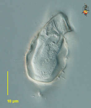
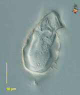
Enchelydium KAHL,1930, species undetermined.Phase contrast.
-
Video showing how this ciiate collected from Cedar Swamp around Woods Hole moves around. Really cute guy.