-
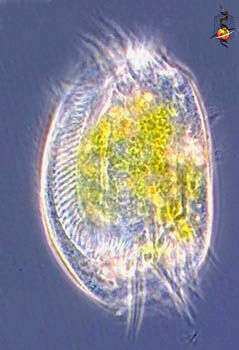
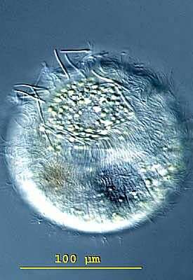
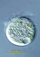
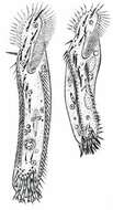
Pelagovasicola (pee-ladge-o-vee-sick-o-la) cinctum is a very fast swimming obovoid ciliate measuring 50 - 180 X 40 - 85 microns. It is common in plankton of lakes and ponds. The body is surrounded by 5-7 distinct ciliary girdles. The posterior fifth of the cell is unciliated. The contractile vacuole lies in the posterior end and has about 20 radial collecting channels. The macronucleus is kidney-shaped and lies in the mid-body. Extrusomes are arranged in the margin of the oral dome, occasionally extruded as bundles of fine filaments. This free-swimming specimen was collected in the plankton of a bog pond near Konstanz, Germany. 115 X 92 microns. Differential interference contrast.
-
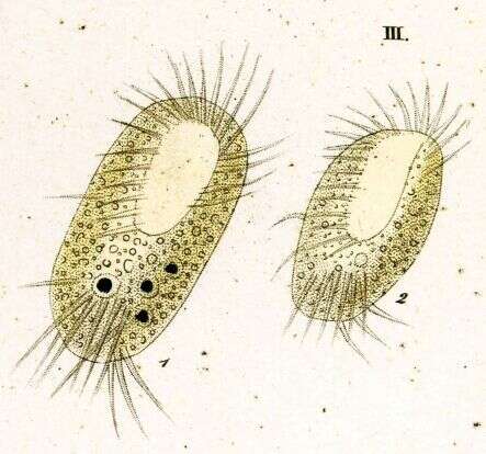
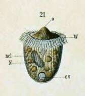
Originally described by Schewiakoff under the name Didinium balbianii. The "rear offspring" (i.e. opisthe) of a recent division. cv--Contractile vacuole N--Macronucleus ncl--Micronucleus o--Mouth W--Ciliated ring
-
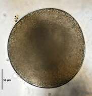
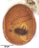
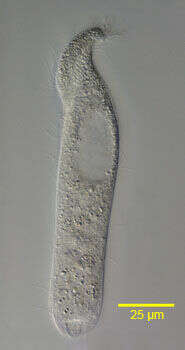
Resting cyst of Urostyla grandis (EHRENBERG,1830). Brightfield.
-
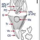
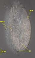
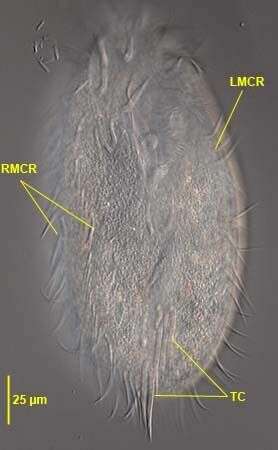
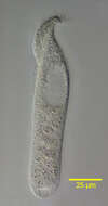
Ventral view of Pleurotricha lanceolata (EHRENBERG,1835) STEIN, 1859. RMCR= two right marginal cirral rows. LMCR=single left marginal cirral row.T=transverse cirri in 2 groups (3+2).Collected from a flood-irrigated grass lawn in Boise,Idaho. May 2008. DIC.
-
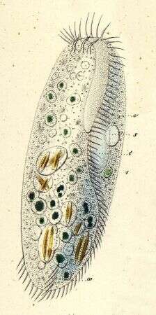
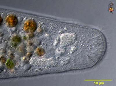
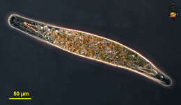
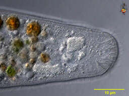
Homalozoon, a elongate ribbon-like predatory ciliate. The body is truncated (cut) off at the front end where the mouth is located, and pointed posteriorly. It has rows of cilia mostly on the ventral side, it glides over the substrate, sometimes contracting. Feeds on detritus and other protists. Phase contrast micrograph.
-
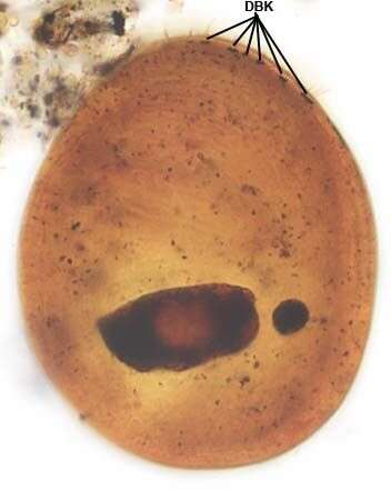
Dorsal infraciliature of Gastronauta derouxi (FOISSNER & BLATTERER, 1992).DBK=dorsal brush dikinetids. Collected from an ephemeral puddle on a grass lawn in Boise, Idaho. July 2007. Stained by the silver carbonate technique (Foissner,W. Europ. J. Protistol.27:313-330;1991).Brightfield.
-
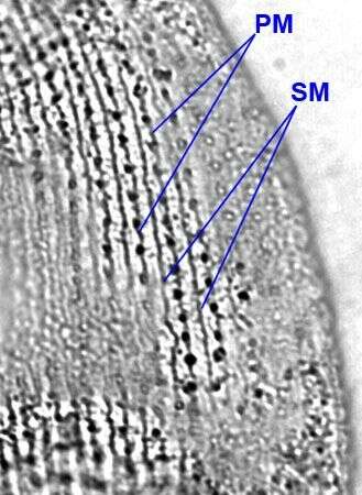
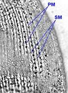
Silverline system of Colpidium kleini (FOISSNER, 1969).There is a single secondary meridian (SM) between each pair of primary meridians (PM).This feature distinguishes C. kleini from the larger C. colpoda whose silverline system shows two secondary meridians between pairs of primary meridians.Stained by the dry silver nitrate technique (see Foissner, W. Europ. J. Protistol., 27:313-330;1991).Brightfield.
-
Image from Li et al., 2010. Acta Protozoologica, 49: 195-212.
-
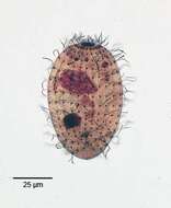
A species of the marine hypotrich genus, Diophrys (DUJARDIN,1840). The genus contains many species. Collected from a commercial marine aquarium in Boise, Idaho. Phase contrast.
-
Euplotes (you-p-low-tees) is a hypotrich ciliate. The hypotrichs form part of the spirotrichs, and most have a large adoral zone of membranelles curving around the front of the cells and terminating at the cytostome on the ventral surface. They are called hypotrichs because the cilia that are used for locomotion are located mostly on the ventral side of the cilia. The cilia are clustered into aggregates called cirri. Euplotids feed on suspended particles such as bacteria and algae. Common. Phase contrast.
-
-
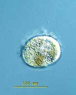
Pelagovasicola (pee-ladge-o-vee-sick-o-la) cinctum is a very fast swimming obovoid ciliate measuring 50 - 180 X 40 - 85 microns. It is common in plankton of lakes and ponds. The body is surrounded by 5-7 distinct ciliary girdles. The posterior fifth of the cell is unciliated. The contractile vacuole lies in the posterior end and has about 20 radial collecting channels. The macronucleus is kidney-shaped and lies in the mid-body. Extrusomes are arranged in the margin of the oral dome, occasionally extruded as bundles of fine filaments. This slightly squashed specimen was collected in the plankton of a bog pond near Konstanz, Germany, and this images emphasizes the radial collecting channels of the contractile vacuole. Differential interference contrast.
-
Originally described by Ehrenberg under the name Stylonychia appendiculata.
-
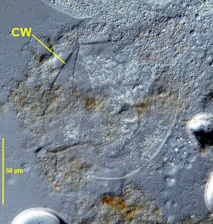
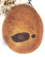
Ruptured resting cyst of Urostyla grandis (EHRENBERG,1830) showing very thin cyst wall (CW). DIC.
-
Originally described by Ehrenberg under the name Stylonychia lanceolata.
-
Homalozoon, a elongate ribbon-like predatory ciliate. The body is truncated (cut) off at the front end where the mouth is located, and pointed posteriorly. It has rows of cilia mostly on the ventral side, it glides over the substrate, sometimes contracting. Feeds on detritus and other protists. This image is of the mouth and shows the needle-like extrusomes lying under the cell surface - these are used to kill and capture prey. Behind the extrusomes is adense refractile mass that is involved in the feeding process. Differential interference contrast.
-
Ventral infraciliature of Gastronauta derouxi (FOISSNER & BLATTERER, 1992).Collected from an ephemeral puddle on a grass lawn in Boise, Idaho. July 2007. Stained by the silver carbonate technique (Foissner,W. Europ. J. Protistol.27:313-330;1991).Brightfield.
-
Right side view of Metopus palaeformis (Kahl, 1927).Synonyms probably include Tesnospira alba (Jankowski,1964),M. hyalinus (Kahl,19270 and M. tenuis (Kahl,1927) among others.Morphology is highly variable probably explaining the large number of synonyms. The cell is flask-shaped to elongate (as in this example).The anterior end is twisted to the left resulting in a rounded lip that overhangs the peristome.The spiral peristome is bordered on the left by an adoral zone of membranelles and on the right by five closely spaced kineties,the "perizonal stripe".Just to the right of the posterior termination of the AZM is a short, inconspicuous undulating membrane(usually visible only in silver-stained preparations).The The right somatic kineties parallel the peristome anteriorly and the left somatic kineties terminate at the margin of the peristome.There is no long tuft of caudal cilia. The prominent ellipsoid macronucleus and adjacent micronucleus are in the anterior half. The contractile vacuole is at the posterior end.The cytoplasm contains endosymbiotic methanogenic bacilli (not seen here).There is an aggregate of brown refractile granules at the anterior end typical of the metopid ciliates.Collected from the bottom sediments of an organically enriched rain pool with abundant decaying grass contaminated by Canada goose (Branta canadensis) droppings.Boise, Idaho. January 2006. DIC.
-
Image by Li et al., 2010. Acta Protozoologica, 49: 195-212.
-
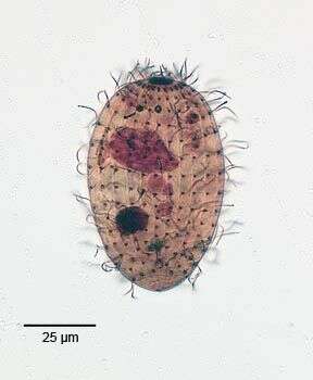
Aspidisca (as-pid-isk-a) is a hypotrich ciliate, and identifiable as a hypotrich because it uses clumps of cilia (cirri) on the ventral surface to walk over the substrate. Hypotrichs are part of the polyhymenophora, and usually feed using an extensive adoral zone of membranelles which extends from the front of the cell to a mouth in the posterior ventral part of the cell. However, in Aspidisca, the AZM has been greatly reduced and forms a kind of scrubbing brush on the ventral surface . Differential interference contrast.
-
-
-
Pelagovasicola (pee-ladge-o-vee-sick-o-la) cinctum is a very fast swimming obovoid ciliate measuring 50 - 180 X 40 - 85 microns. It is common in plankton of lakes and ponds. The body is surrounded by 5-7 distinct ciliary girdles. The posterior fifth of the cell is unciliated. The contractile vacuole lies in the posterior end and has about 20 radial collecting channels. The macronucleus is kidney-shaped and lies in the mid-body. Extrusomes are arranged in the margin of the oral dome, occasionally extruded as bundles of fine filaments. This slightly squashed specimen was collected in the plankton of a bog pond near Konstanz, Germany, and this images emphasizes the extruded extrusomes at the margin of the oral dome. Differential interference contrast.
-
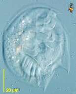
Infraciliature of the planktonic protstomatid ciliate, Apsiktrata gracilis (Penard,1922)Foissner, Berger & Kohmann 1994. Morphologically quite similar to members of the genus Holophrya but lacking a "dorsal brush". The anterior apical cytostome and its circumoral dikinetids is seen here.There is a long caudal cilium in vivo (only its basal body is seen here at the posterior pole). Collected from a freshwater pond near Boise, Idaho. Silver carbonate stain (see Foissner, W. Europ. J. Protistol., 27:313-330;1991). Brightfield