-
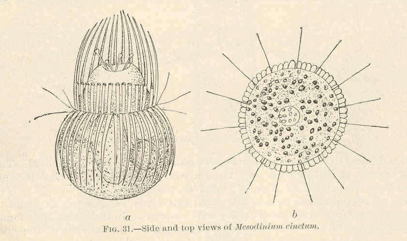
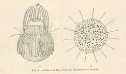
Mesodinium cinctum--Side and top views.
-
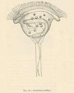
Vorticella patellina.
-
-
Phase contrast micrograph of living cell.
-
Infraciliature of a middle divider of Monodinium balbianii (FABRE_DOMERGUE,1888). The ciliary girdles of the opisthe and proter are visible here. Densely stained extrusomes are also visible.Stained by the silver carbonate technique (see Foissner, W. Europ. J. Protistol., 27:313-330;1991). Brightfield.
-
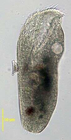
in vivo portrait (ventral view) of the large hypotrich ciliate, Urostyla grandis (EHRENBERG,1830). Collected from a freshwater canal near Boise, Idaho.Brightfield.
-
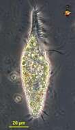
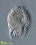
Trachelostyla (track-ell-owe-stike-a) (tentative identification) is one of a large number of stichotrichine hypotrich ciliates, and the genera can only be properly distinguished by careful mapping of the the distribution of the cirri - an exercise which requires special preparation of the cell. Trachelostyla is one of several genera which can have a narrowed front end bearing the adoral zone of membranelles. Phase contrast.
-
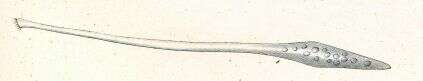
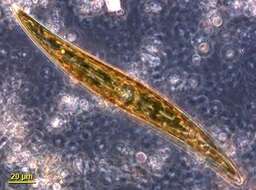
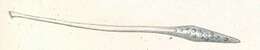
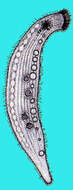
Originally described by Ehrenberg under the name Trachelocerca olor.
-
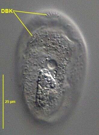
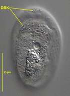
Dorsal view of Gastronauta membranaceus (Engelmann in Bütschli,1889). DBK= dorsal brush kineties. The bipartite dorsal brush is one of the distinguishing characteristics of this species. G. derouxi has a long anterior marginal dorsal brush row of dikinetids. From a freshwater pond near Boise, Idaho. DIC.
-

Right lateral infraciliature of the hymenostome ciliate Colpidium kleini (Foissner, 1969). C. kleini is very similar in overall appearance to C. colpoda although usually more slender and with fewer somatic kineties. The cytostome is in the anterior 1/4 of the cell. There is a curved paraoral membrane along the convex right margin of the cytostome. The left margin is slightly concave. There are three adoral membranelles. There are 32 to 44 somatic kineties. The kineties to the right and left of the oral aperture meet at a curved preoral suture. The right somatic kineties bend leftward at the level of the cytostome. There is an anterior apical area bare of cilia. There are rows of inconspicuous mucocysts between the somatic kineties. The ellipsoid macronucleus and adjacent micronucleus are centrally located. The single contractile vacuole is located in the midbody with a single excretory pore on the right surface. The feature most clearly distinguishing Colpidium kleini from C. coploda is the silverline system (as demonstrated by silver nitrate staining).Stained by the silver carbonate technic (see Foissner, W.Europ. J. Protistol.27,313-330;1991). Collected from an organically enriched freshwater pond near Boise, Idaho.Brightfield.
-
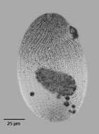
Gonostomum affine (Stein, 1859) Sterki, 1878. Non-flooded Petri dish soil sample collected from flood-irrigated lawn in Boise, Idaho, June 2008. Protargol (Wilbert modification). Brightfield.
-
Ventral surface of the hypotricvh ciliate, Uronychia. Isolated from sandy samples taken from Sippiwissett marsh.
-
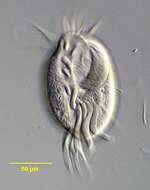
Euplotes (you-ploe-tees) a hypotrich ciliate, with an anterior adoral zone of membranelles forming a collar and lapel leading down to the cytostome which is midway down the cell and on the ventral side. It is a hypotrich ciliate and uses cirri (aggregates of ciliate, sometimes as many as 50 or more) to walk against the substrate. Identification is approximate as the full identification requires that cells be silver-stained and the location of each cirrus be mapped out. DIfferential interference contrast.
-
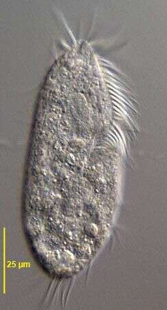
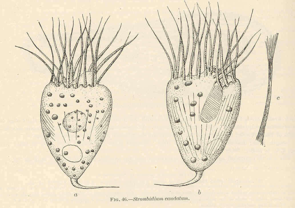
Strombidium caudalum.
-
-
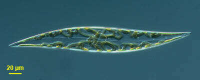
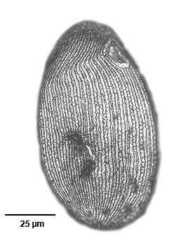
This pennate diatom was found in a plankton tow from Nantucket Sound off Martha's Vineyard - Massachusetts, USA. Image by Jeffrey Cole and Micah Dunthorn.
-
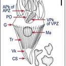
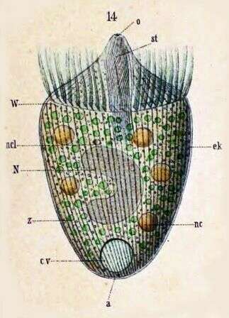
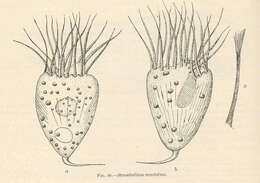
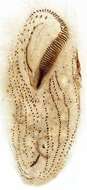
Originally described by Schewiakoff under the name Didinium balbianii. An individual with weakly projecting mouth cone, traveling backwards. a--Anus cv--Contractile vacuole ek--Ectoplasm N--Macronucleus ncl--Micronucleus o--Mouth st--Cytopharyngeal basket W--Ciliated ring z -- Zoochlorellae
-
Ventral infraciliature of the large hypotrich ciliate, Urostyla grandis (EHRENBERG,1830). Collected from a freshwater canal near Boise, Idaho.Stained by the protargol A technique (see Foissner, W. Europ. J. Protistol., 27:313-330;1991).Brightfield.
-
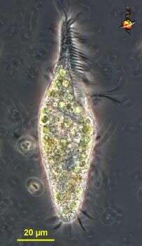
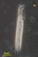
Trachelostyla (track-ell-owe-style-a), a hypotrich ciliate. With a narrowed anterior end to the body, with an adoral zone of membranelles. Phase contrast micrograph.
-
-
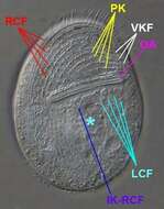
Ventral view (in vivo) of Gastronauta derouxi (FOISSNER & BLATTERER, 1992). OA=oral large transverse oral aperture. RCF=right ventral somatic ciliary field. LCF= left ventral somatic ciliary field. PK=preoral kineties. VKF=vertical kinety fragments.IK-RCF=anteriorly curved innermost kinety of right ciliary field. *= unciliated postoral area between right and left somatic ciliary fields.Collected from an ephemeral puddle on a grass lawn in Boise, Idaho. July 2007.DIC
-
Right lateral view of the silverline system of the hymenostome ciliate Colpidium kleini (Foissner, 1969). C. kleini is very similar in overall appearance to C. colpoda although usually more slender and with fewer somatic kineties. The cytostome is in the anterior 1/4 of the cell. There is a curved paraoral membrane along the convex right margin of the cytostome. The left margin is slightly concave. There are three adoral membranelles. There are 32 to 44 somatic kineties. The kineties to the right and left of the oral aperture meet at a curved preoral suture.The right somatic kineties bend leftward at the level of the cytostome. There is an anterior apical area bare of cilia. There are rows of inconspicuous mucocysts between the somatic kineties. The ellipsoid macronucleus and adjacent micronucleus are centrally located. The single contractile vacuole is located in the midbody with a single excretory pore on the right surface. The feature most clearly distinguishing Colpidium kleini from C. coploda is the silverline system. In C. kleini there is only one secondary meridian (silverline) between two primary meridians (primary meridians correspond to somatic kineties). In some cases short segments of the secondary meridians may be duplicated. Short transverse L or T-shaped branches arise from both primary and secondary meridians at irregular intervals.Stained by the dry silver nitrate technic (see Foissner, W.Europ. J. Protistol.27,313-330;1991). Collected from an organically enriched freshwater pond near Boise, Idaho. Brightfield. Black and white.
-
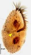
Ventral infraciliature of the oxytrichid,Gonostomum strenuum (ENGELMANN, 1862) STERKI, 1878. G. strenuum differs from G. affine by the greater number of frontoterminal (4-6 vs 2) and frontoventral cirri. In G. strenuum the last frontoventral cirrus is just posterior to the peristome (yellow arrowhead).Collected from a non-flooded Petri dish culture of soil from a park lawn in Boise, Idaho. January 2007.Stained by the protargol technique [Wilbert modification] (see Foissner, W. Europ. J. Protistol., 27:313-330;1991).Brightfield.
-
A species of the marine hypotrich genus, Diophrys (DUJARDIN,1840). The genus contains many species. Collected from a commercial marine aquarium in Boise, Idaho. DIC.