-
-
-
-
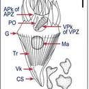
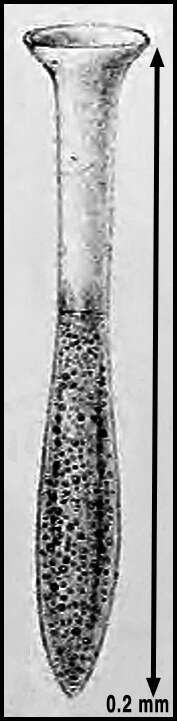
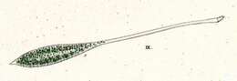
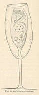
Drawing from Claparde & Lachmann 1858 (Plate 8, figure 5) of the species now known as Steenstrupiella steenstrupii as "Tintinnus steenstrupii". The scale bar reflects the text description (pg 200) as about 0.2 mm in overall length. The species (and now genus) was presumably named after the Danish biologist Japetus Steenstrup (1813-1897).
-
-
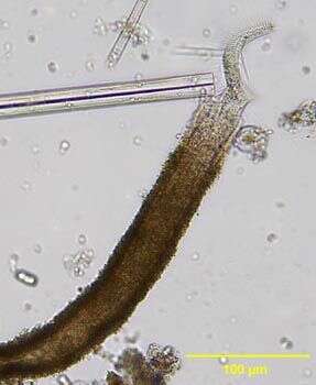
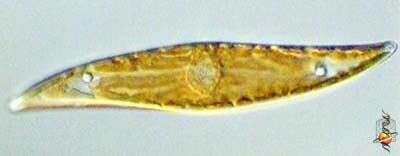
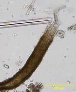
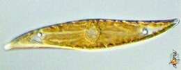
Pleurosigma (ploo-row-sig-ma) and Gyrosigma are two rather similar genera of sigmoid-shaped pennate diatoms found in intertidal sediments, salt marshes and so on. The nucleus is located at the centre of the cell. The plastids contain chlorophylls a and c which gives the yellowy-brown colour. . Pleuosigma is distinguished in part by the angled pattern of marks on the valve of the frustule. Differential interference contrast.
-
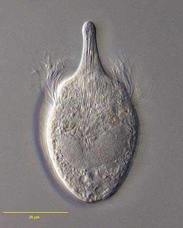
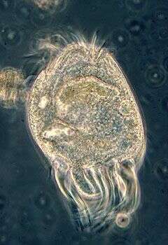
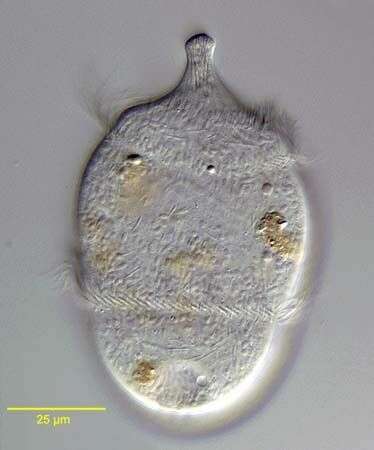
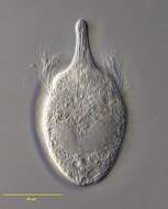
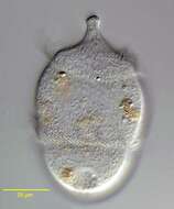
Portrait of the haptorid ciliate, Monodinium balbianii (FABRE-DOMERGUE,1888). The body is a stout cup shape, sometimes more narrowly rounded posteriorly. The oral aperture is at the end of a prominent cone-shaped proboscis that protrudes from the center of the truncate anterior end. The proboscis contains many long toxicysts. There are 50-100 rows of basal bodies but only a narrow circumferential band of these forms the single anterior ciliary girdle. The midbody macronucleus is horseshoe-shaped (as seen in section here) or reniform. There is a single spherical micronucleus (not seen here). The contractile vacuole is posterior. Swims very rapidly, rotating on long axis. Feeds on small flagellates and other ciliates. Collected from organically enriched freshwater pond near Boise, Idaho in September 2003. DIC optics.
-
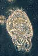
Eutintinnus (you-tin-tin-us), one of the tintinnid ciliates. These are mostly marine choreotrichs in which the cell is located within an open lorica which it drags around while it swims. The different genera and species are mostly distinguished by the different appearances of the lorica. This genus has a conical lorica which is open at both ends. With an adoral zone of membranelles located around the top end of the cell. Phase contrast.
-
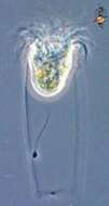
Portrait of Chaetospira, a loricate hypotrich ciliate. Elongate anterior end with prominent adoral zone of membranelles has a corkscrew configuration, distinguishing this genus from the similar Stichotricha. Usually only anterior portion protrudes and organism quickly retracts completely into lorica when disturbed. From freshwater pond near Boise, Idaho. Brightfield.
-
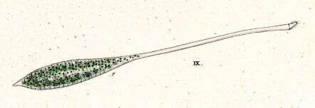
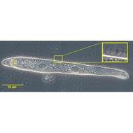
Originally described by Ehrenberg under the name Trachelocerca viridis.
-

Gastronauta membranaceus (Engelmann in Bütschli,1889), a hypostome ciliate, distinguished by its long transversely oriented cytostome. The cytostome lacks trichites. The body is ovoid in outline and strongly dorsoventrally flattened. Ciliature is restricted to the ventral surface except for two short dorsal kineties anteriorly. A few somatic kineties run uninterrupted to the right of the cytostome arching around the anterior of the cell. Several right somatic kineties are interrupted by the cytostome. The left somatic kineties terminate at the cytostome. A single kinety runs around the circumference of the cytostome. An unciliated bare are overlies the region of the macronucleus posterior to the cytostome. The macronucleus is oblong and heteromerous (i.e. containing areas with markedly differing RNA and DNA contents resulting in irregular staining and optical characteristics). The single micronucleus is quite prominent. Two contractile vacuoles are present, one in the anterior half and one posteriorly. Gastronauta feeds mainly on diatoms. From a freshwater pond near Boise, Idaho. Phase contrast illumination.
-
Luporinophrys micelae (FOISSNER,2005). Collected from an ephemeral puddle on a flood-irrigated grass lawn in Boise, Idaho, 2007.Phase contrast.
-
Lugol's fixed specimen
-
Marine species, very well developed cirri. Phase contrast micrograph of living cell.
-
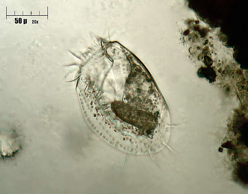
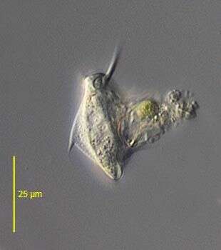
Lateral view of the hypotrich ciliate, Aspidisca turrita (Ehrenberg, 1831) Claparède & Lachmann, 1858. The thorn-like dorsal process is clearly seen here (to viewer's left). The first ventral cirrus is also visible projecting anteriorly. Collected from a freshwater pond near Boise, Idaho. June 2005.DIC
-
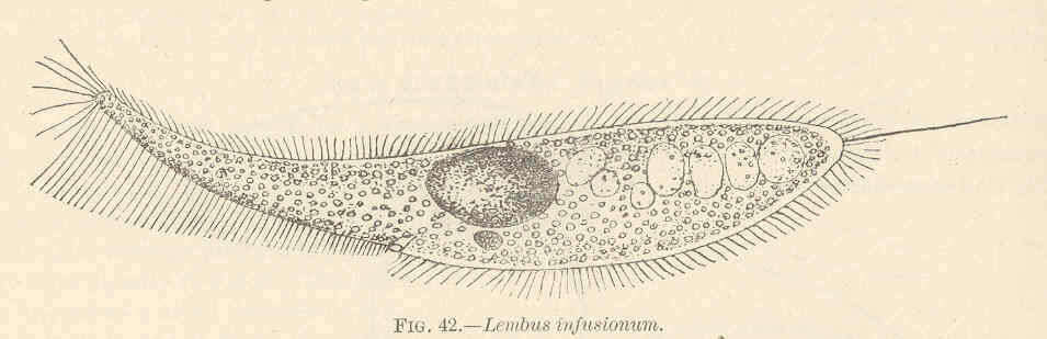
Lembus infusionum.
-
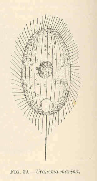
Uronema marina.
-
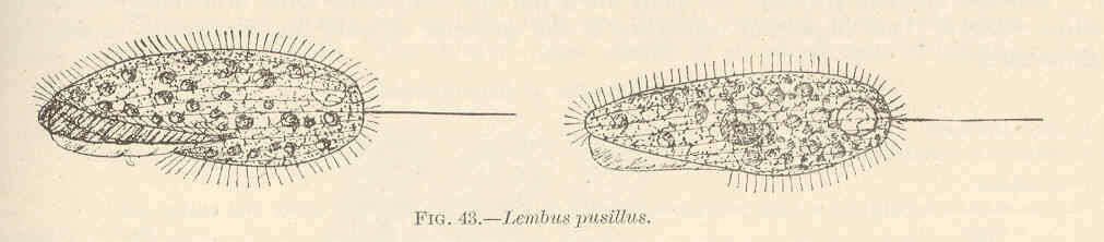
Lembus pusillus.
-
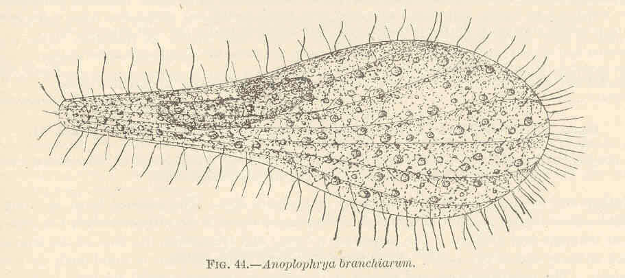
Anoplophrya.
-
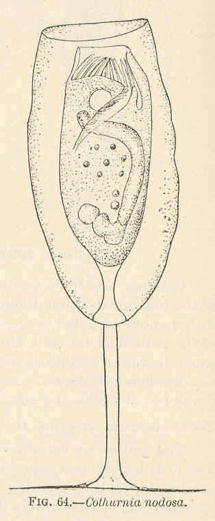
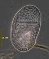
Cothurnia nodosa.
-
-
-
Pleurosigma (ploo-row-sig-ma) and Gyrosigma are two rather similar genera of sigmoid-shaped pennate diatoms found in intertidal sediments, salt marshes and so on. The nucleus is located at the centre of the cell. The plastids contain chlorophylls a and c which gives the yellowy-brown colour. . Pleuosigma is distinguished in part by the angled pattern of marks on the valve of the frustule. Differential interference contrast.
-
Portrait of Monodinium balbianii (FABRE-DOMERGUE,1888) in mid-division. Dividers are easily confused with Didinium species but may be distinguished by the furrow associated with the ciliary girdle of the opisthe (posterior daughter cell). Stomatogenesis is telokinetal (i.e. new oral ciliature develops from anterior ciliary rows of opisthe). Collected from organically enriched freshwater pond near Boise, Idaho in September 2003. DIC optics.