-
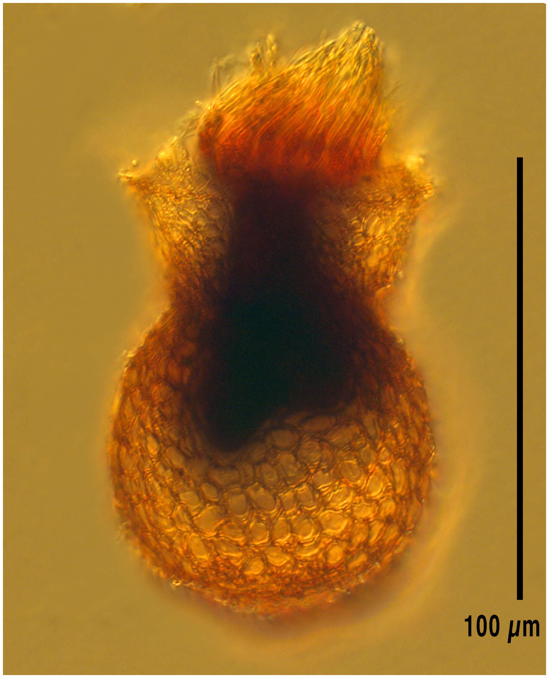
From the Bay of Villefranche in December 2013, lugol's-fixed specimen, Z-stack of images made using a 60x objective and DIC optics.
-
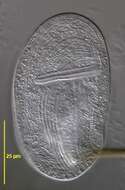
Specimen lugol's-fixed from the Ionian Sea.
-
-
-
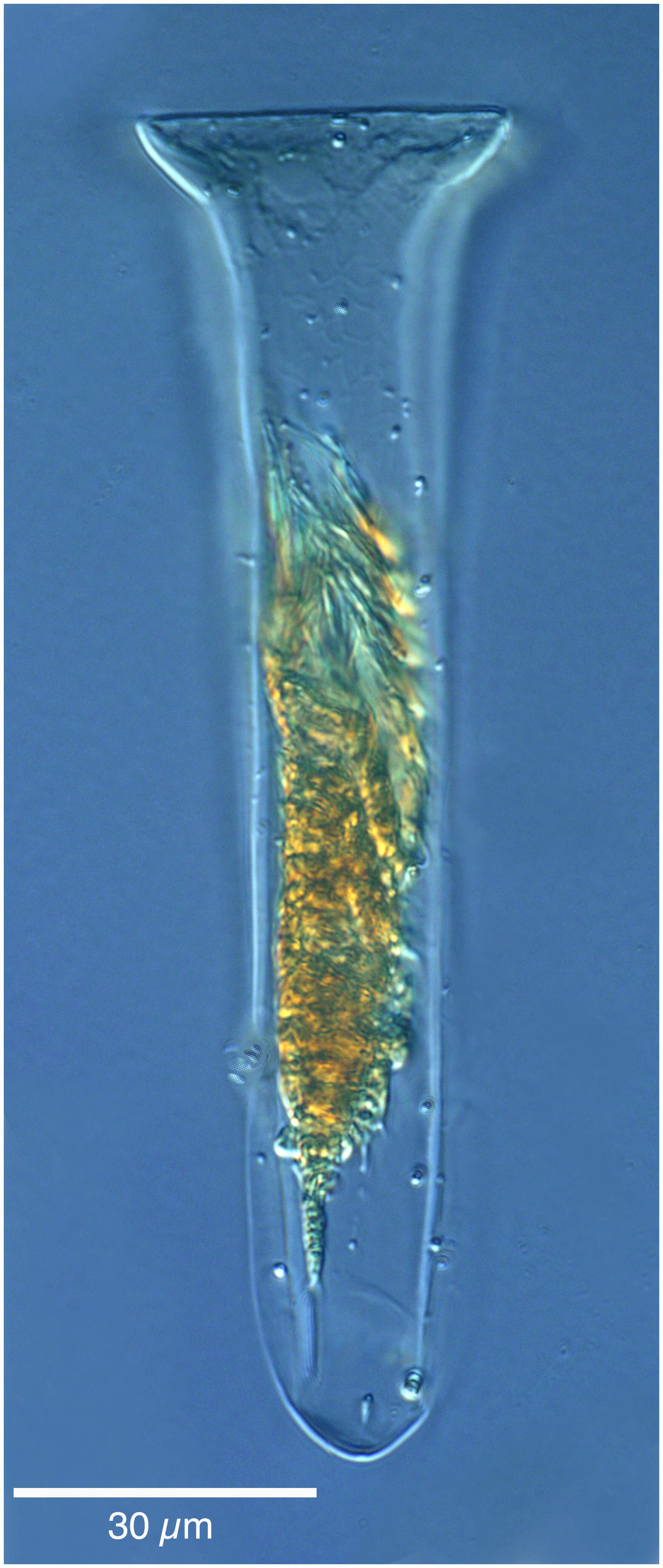
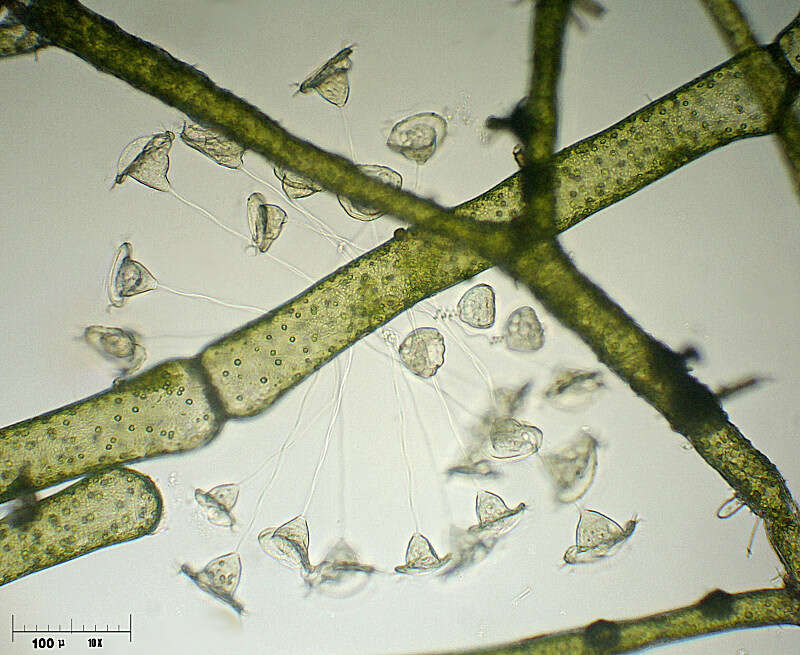
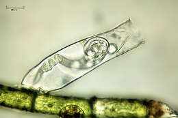
Codonaria cistellula from the Bay of Villefranche in October 2014. Z-stack of images made using a 20x objective and DIC optics, lugol's-fixed specimen.
-
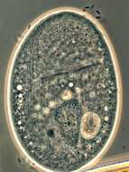
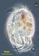
A ciliated protozoon, from Lake Mono, California.
-
-
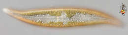
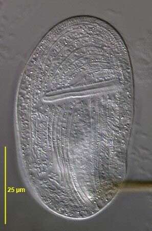
Pleurosigma (ploo-row-sig-ma) and Gyrosigma are two rather similar genera of sigmoid-shaped pennate diatoms found in intertidal sediments, salt marshes and so on. The nucleus is located at the centre of the cell. The plastids contain chlorophylls a and c which gives the yellowy-brown colour. This picture is taken of the surface of one of the valves and shows the raphe that is used in locomotion, and shows the plastids. Refractile globules are said to be the storage products from excessive photosynthesis. Pleuosigma is distinguished in part by the angled pattern of marks on the valve of the frustule. Differential interference contrast.
-
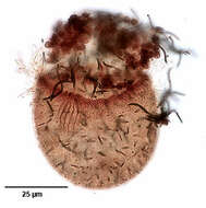
Dorsal infraciliature of the haptorid ciliate, Monodinium balbianii (FABRE-DOMERGUE, 1888). The dorsal side is distinguished by the five parallel rows of clavate (club-shaped) cilia (the dorsal brush) just posterior to the anterior wreath of ciliated basal bodies. The longitudinal files of somatic kinetosomes (not seen well here) are unciliated except for the dorsal brush and the obliquely inclined closely spaced ciliated basal bodies that form the ciliary girdle. Darkly stained extrusomes are visible here. Collected from a freshwater pond near Boise, Idaho March 2005. Stained by the silver carbonate technique (see Foissner, W. Europ. J. Protistol., 27:313-330;1991). Brightfield.
-
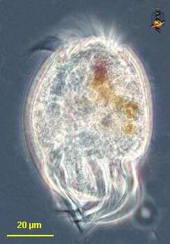
Eutintinnus (you-tin-tin-us), one of the tintinnid ciliates. These are mostly marine choreotrichs in which the cell is located within an open lorica which it drags around while it swims. The different genera and species are mostly distinguished by the different appearances of the lorica. This genus has a conical lorica which is open at both ends. With an adoral zone of membranelles located around the top end of the cell. Differential interference contrast.
-
-
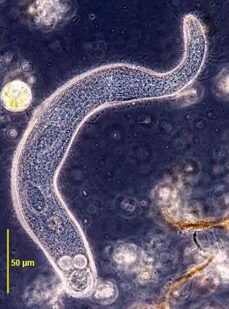
Originally described by Ehrenberg under the name Trachelocerca viridis.
-
Gastronauta membranaceus (Engelmann in Bütschli,1889), a hypostome ciliate, distinguished by its long transversely oriented cytostome. The cytostome lacks trichites. The body is ovoid in outline and strongly dorsoventrally flattened. Ciliature is restricted to the ventral surface except for two short dorsal kineties anteriorly. A few somatic kineties run uninterrupted to the right of the cytostome arching around the anterior of the cell. Several right somatic kineties are interrupted by the cytostome. The left somatic kineties terminate at the cytostome. A single kinety runs around the circumference of the cytostome. An unciliated bare are overlies the region of the macronucleus posterior to the cytostome. The macronucleus is oblong and heteromerous (i.e. containing areas with markedly differing RNA and DNA contents resulting in irregular staining and optical characteristics). The single micronucleus is quite prominent. Two contractile vacuoles are present, one in the anterior half and one posteriorly. Gastronauta feeds mainly on diatoms. From a freshwater pond near Boise, Idaho. DIC. This image was taken by William Bourland. He now uses a Zeiss Axioskop 2 with a Spot Insight CCD camera (Diagnostic Instruments).
-
Luporinophrys micelae (FOISSNER,2005). Collected from an ephemeral puddle on a flood-irrigated grass lawn in Boise, Idaho, 2007.Phase contrast.
-
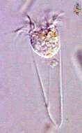
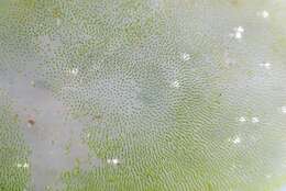
Lugol's fixed specimen
-
Uronychia is a hypotrich ciliate. As such it has an adoral zone of membranelles for feeding, and these extend around the anterior end of the cell. Like other hypotrichs, motile cilia are clustered into cirri on the ventral surface. Those of Diophrys are very strongly developed. Phase contrast.
-
-
Almind Sø, Jylland, Danmark
-
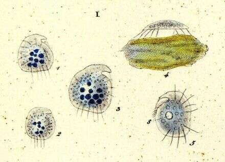
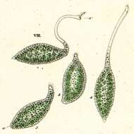
Loxophyllum setigerum, var. armatum. a,b,c: ventral, dorsal, and lateral aspects.
-
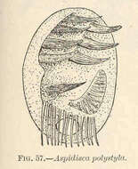
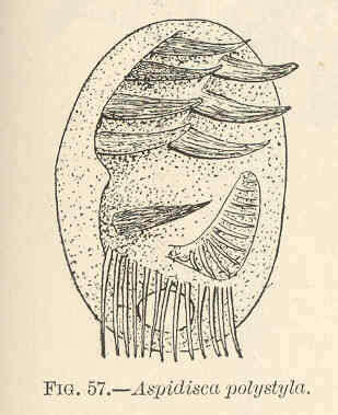
Aspidisca polystyla.
-
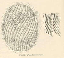
Nassula microstoma.
-
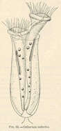
Cothurnia imberbis.
-
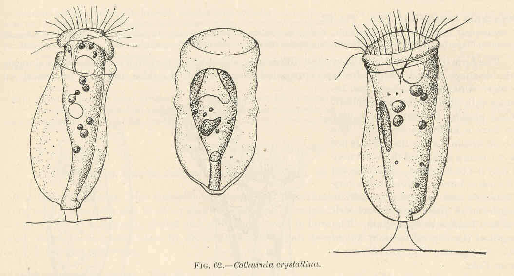
Cothurnia crystallini.
-
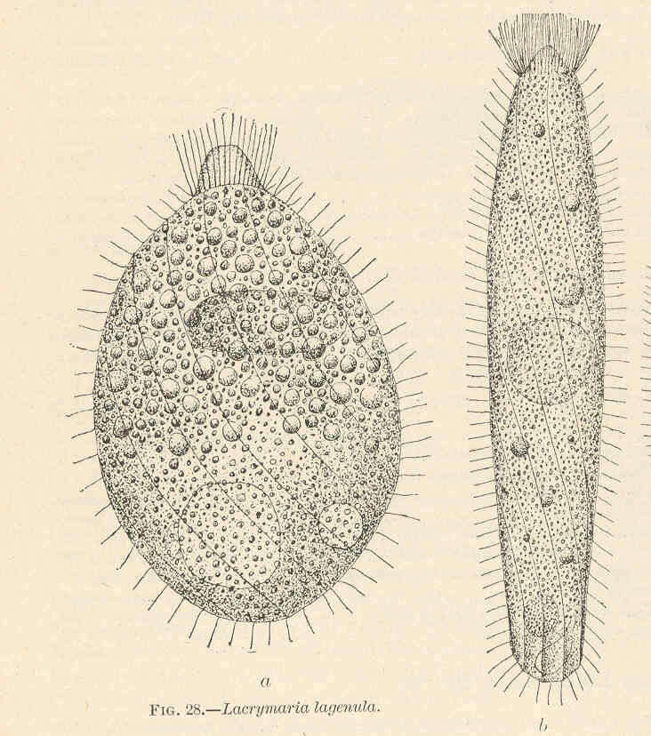
Lacrymaria lagenula.