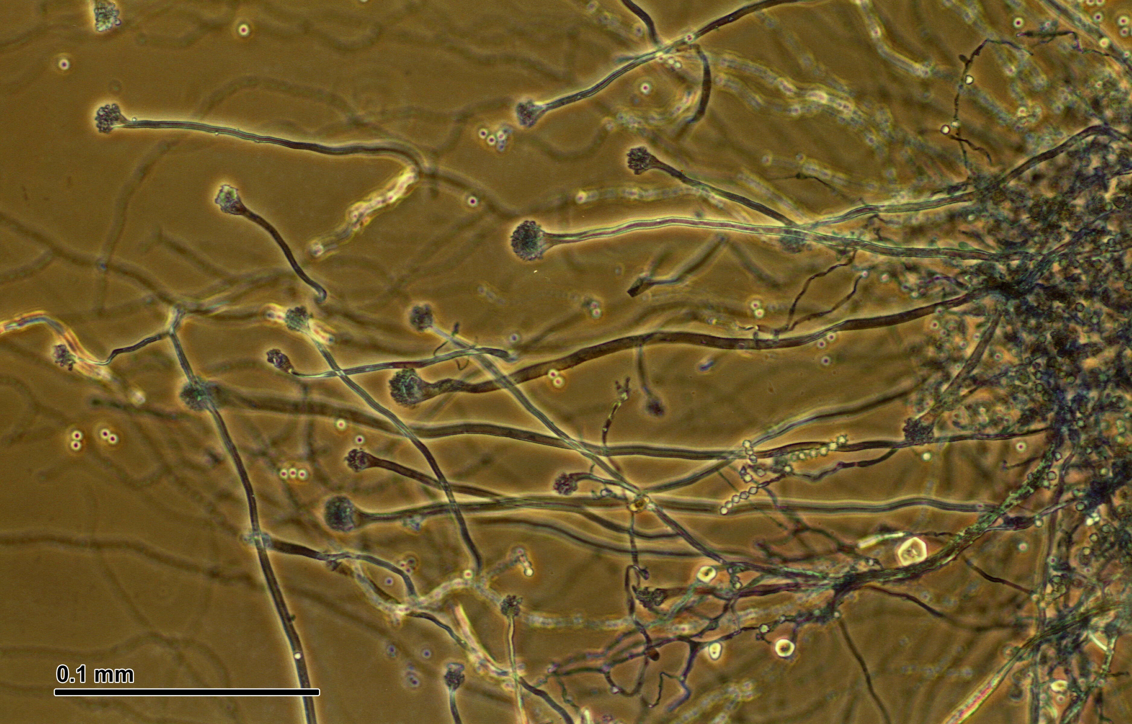
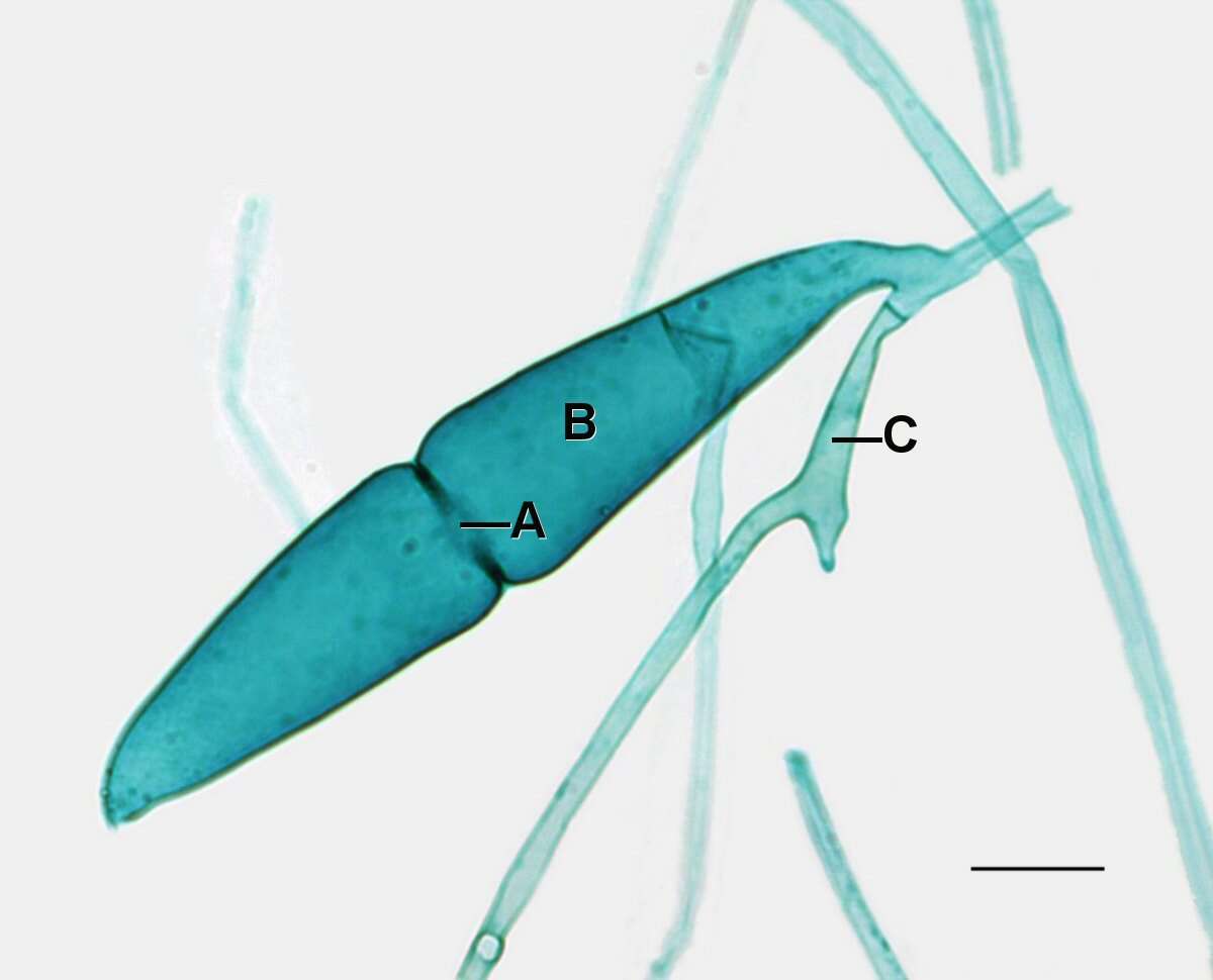
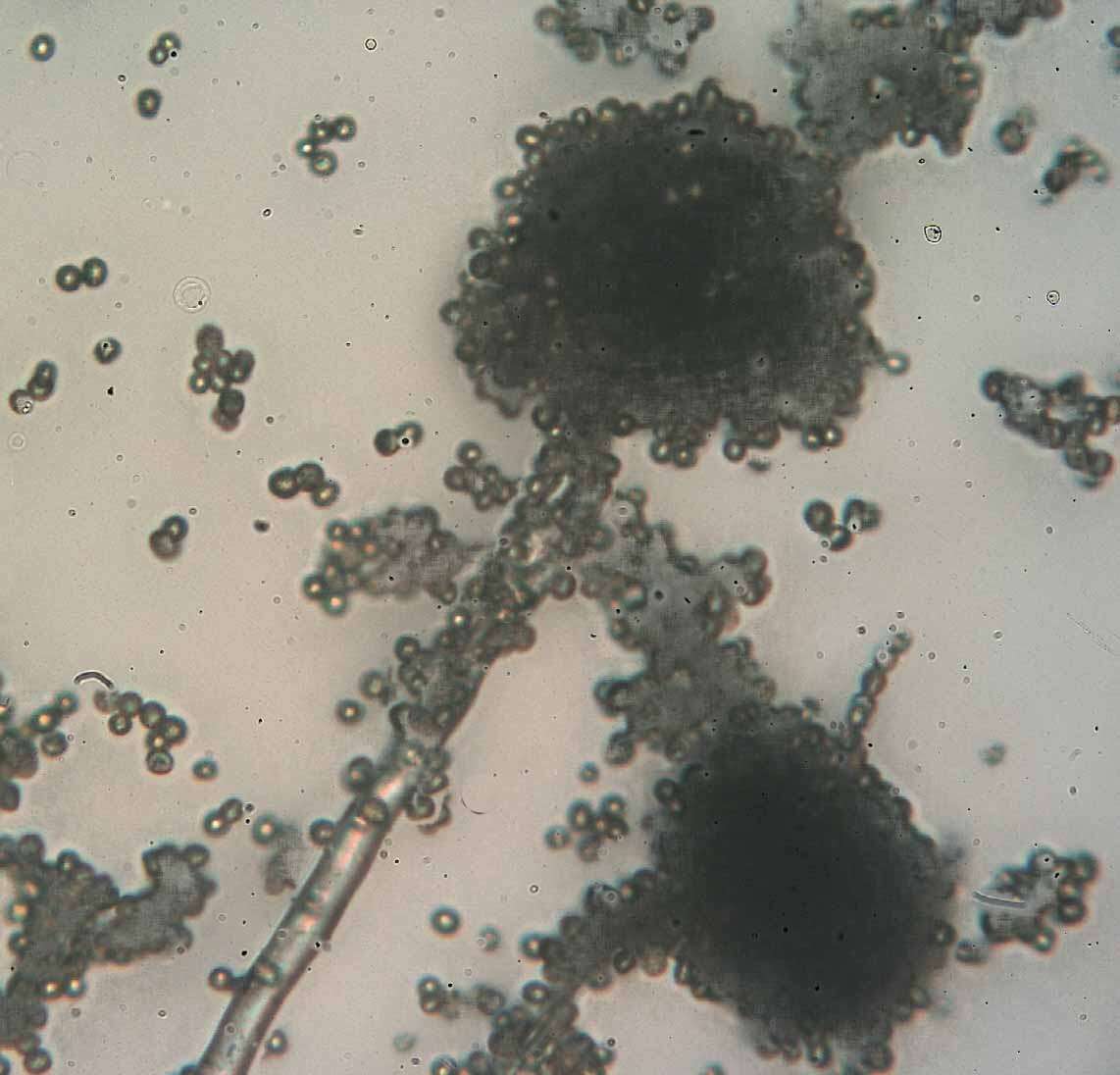
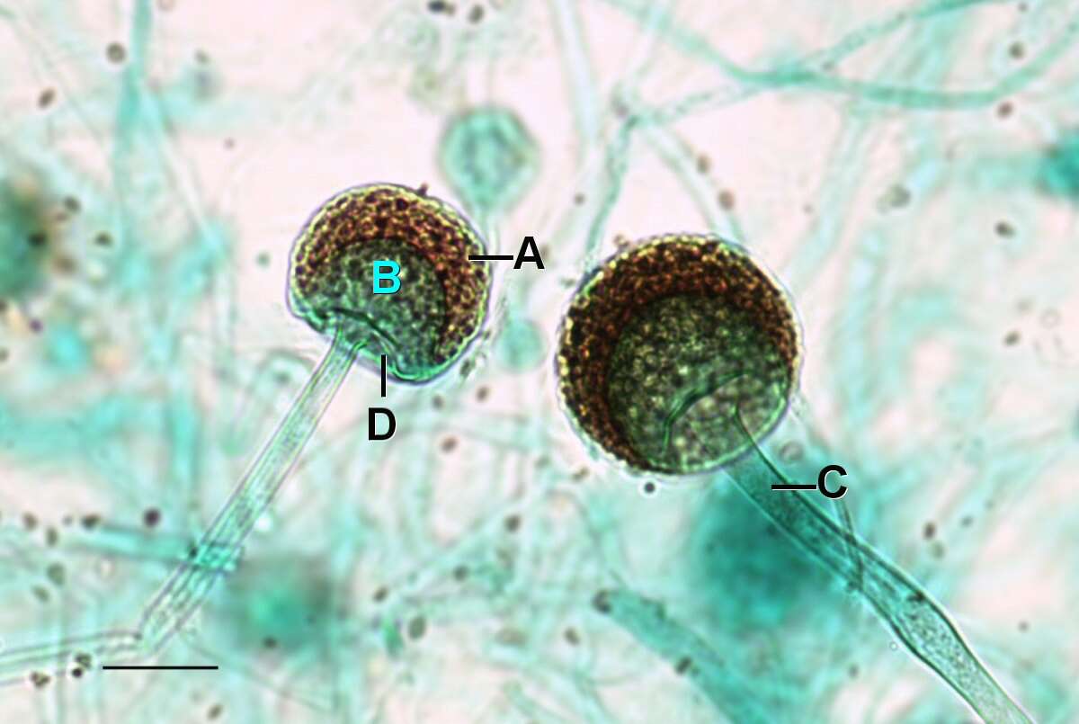
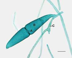
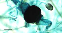
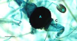
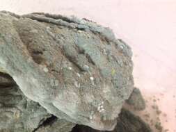
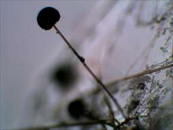
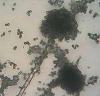
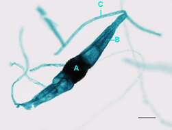
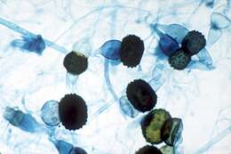
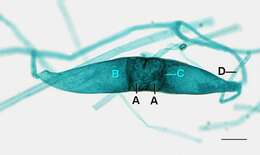
Description: English: Light microscopy of Rhizopus showing a close up view of two sporangium of Rhizopus where the differentiation between the spores and columella can be seen attached to the hypha. A=Sporangium with spores, B=Columella, C=Hypha, D=Apophysis. Scale bar = 0.1mm. Date: 13 May 2014, 11:16:53. Source: Jon Houseman and Matthew Ford. Author:
Jon Houseman. Other versions:
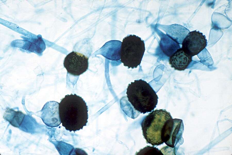
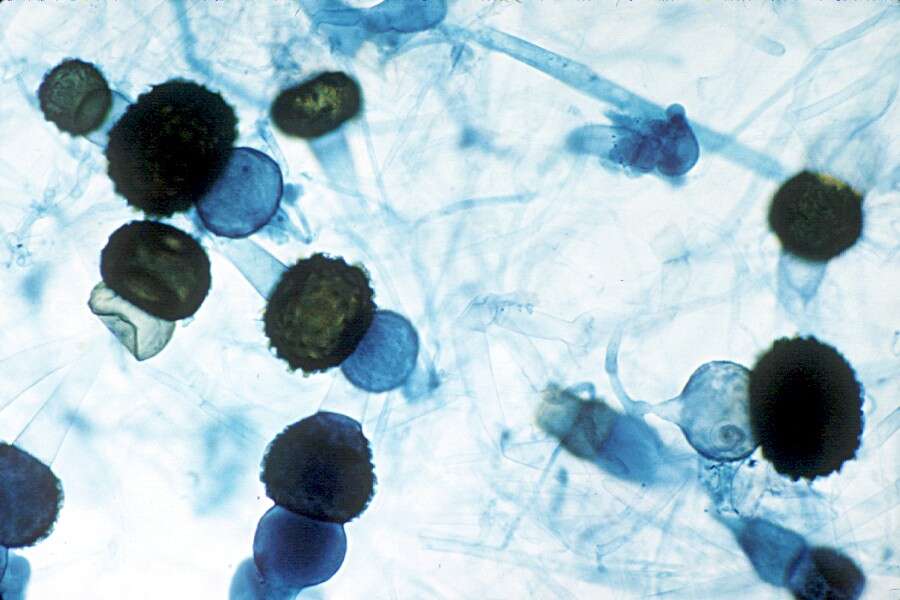
Original (unlabeled).
: This is a
retouched picture, which means that it has been digitally altered from its original version. Modifications: Balance (Color, brightness, and contrast) and adjust background color.


