Wuchereria bancrofti is one of the eight parasitic nematode (roundworm) species that account for most cases of filariasis in humans. This form of filariasis is known as lymphatic filariasis. Furthermore, W. bancrofti is one of the three of these eight species responsible for most of the morbidity attributable to filariasis (the other two being Brugia malayi, which also causes lymphatic filariasis and Onchocerca volvulus, which causes onchocerciasis (river blindness)). Wuchereria bancrofti is encountered in tropical areas worldwide. (Centers for Disease Control Parasites and Health website)
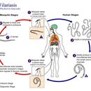
Various species of mosquitoes are known vectors of W. bancrofti filariasis, depending on geographic distribution. Among these are: Culex (C. annulirostris, C. bitaeniorhynchus, C. quinquefasciatus, and C. pipiens); Anopheles (A. arabinensis, A. bancroftii, A. farauti, A. funestus, A. gambiae, A. koliensis, A. melas, A. merus, A. punctulatus and A. wellcomei); Aedes (A. aegypti, A. aquasalis, A. bellator, A. cooki, A. darlingi, A. kochi, A. polynesiensis, A. pseudoscutellaris, A. rotumae, A. scapularis, and A. vigilax); Mansonia (M. pseudotitillans, M. uniformis); and Coquillettidia (C. juxtamansonia). During a blood meal, an infected mosquito introduces third-stage filarial larvae onto the skin of the human host, where they penetrate into the bite wound. They develop in adults that commonly reside in the lymphatics. The female worms measure 80 to 100 mm in length and 0.24 to 0.30 mm in diameter, while the males measure about 40 mm by 0.1 mm. Adults produce microfilariae measuring 244 to 296 μm by 7.5 to 10 μm, which are sheathed and have nocturnal periodicity, except for the South Pacific microfilariae, which lack marked periodicity. The microfilariae migrate into lymph and blood channels moving actively through lymph and blood. A mosquito ingests the microfilariae during a blood meal. After ingestion, the microfilariae lose their sheaths and some of them work their way through the wall of the proventriculus and cardiac portion of the mosquito's midgut and reach the thoracic muscles. There the microfilariae develop into first-stage larvae and subsequently into third-stage infective larvae. The third-stage infective larvae migrate through the hemocoel to the mosquito's prosbocis and can infect another human when the mosquito takes a blood meal. (Centers for Disease Control Parasites and Health website)