Plate 26
Description:
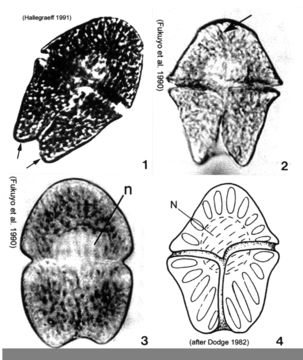
Plate 26. Gymnodinium sanguineum. Figs. 1-3. LM. Cell large, pentagonal, and slightly dorso-ventrally flattened. Cells vary in shape and size. Fig. 1. Ventral view. Epitheca and hypotheca nearly equal in size: epitheca conical, hypotheca bilobed (arrows). Fig. 2. Ventral view. Deep cingulum median, displaced 1-2 times its width. Sulcus deeply notches hypotheca. Apical groove present (arrow). Fig. 3. Cell deeply pigmented; central nucleus (n). Fig. 4. Line drawing. Spindle-shaped chloroplasts radially arranged.
Included On The Following Pages:
This image is not featured in any collections.
Source Information
- license
- cc-publicdomain
- bibliographic citation
- Faust, Maria A. and Rose A. Gulledge. Identifying Harmful Marine Dinoflagellates. Smithsonian Contributions from the United States National Herbarium, volume 42: 1-144 (including 48 plates, 1 figure and 1 table).
- original
- original media file
- visit source
- partner site
- NMNH Marine Dinoflagellates
- ID