"
Description:
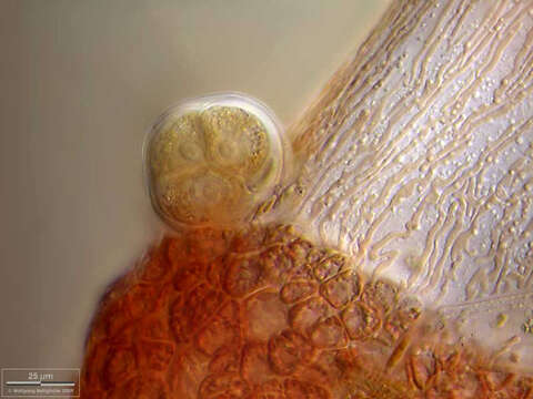
Ceramium diaphanum This detail view of a thallus segment shows the two cell types of the thallus: one tall axial cell (light, with tubular rhodoplasts) and many little reddish cortical cells with their lenticular rhodoplasts. In the tetrasporangium three cells with their nuclei are visible. The scale bar indicates 25 µm. Collected from Bodden, the brackish waters lying between the isles of Hiddensee and Ruegen (German Baltic Sea). This image was taken using Zeiss Universal with Olympus C7070 CCD camera.Image under Creative Commons License V 3.0 (CC BY-NC-SA). Place name: Hiddensee Bodden (Germany) Latitude: 54.582633 Longitude: 13.115051 Diese Detailansicht eines Thallus-Segmenta zeigt Zellen der beiden vorkommenden Typen: eine lange axiale Zelle (hell, mit schlauchförmigen Rhodoplasten) und viele kleine, runde, rötliche, kortikale Zellen mit ihren linsenförmigen Rhodoplasten. Im Tetrasporangium sind drei der vier Zellen mit ihren Kernen sichtbar. Der Messbalken markiert eine Länge von 25 µm. Probe aus dem Hiddenseer Bodden, der Brackwasserfläche zwischen den Inseln Hiddensee und Rügen. Mikrotechnik: Zeiss Universal, Kamera: Olympus C7070. Creative Commons License V 3.0 (CC BY-NC-SA). For permission to use of (high-resolution) images please contact postmaster@protisten.de.
Included On The Following Pages:
- Life (creatures)
- Cellular (cellular organisms)
- Eukaryota (eukaryotes)
- Archaeplastida (plants)
- Rhodophyta (red algae)
- Florideophyceae (Florideae)
- Rhodymeniophycidae
- Ceramiales (An Order of Red Algae)
- Ceramiaceae
- Ceramium
- Ceramium diaphanum
This image is not featured in any collections.
Source Information
- license
- cc-by-nc-sa-3.0
- copyright
- Wolfgang Bettighofer
- creator
- Wolfgang Bettighofer [email]
- original
- original media file
- visit source
- partner site
- protisten.de
- ID