Image of horseshoe worms
Description:
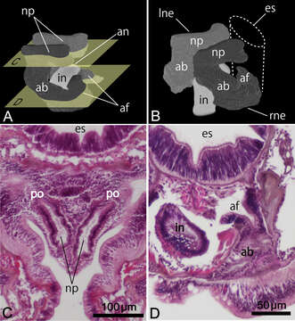
Figure 5.Reconstructed three-dimensional images and transverse sections of the nephridium of Phoronis ijimai Oka, 1897, from NSMT-Te 886 (A, B, D) and NSMT-Te 884 (C). A Lateral view, showing the long nephridial papillae above the anus B dorsolateral view, showing the offset arrangement of the nephridia, with the curved ascending branch and large anal funnel extending toward the esophagus C transverse section through the nephridial papilla, showing the nephridiopore D transverse section through the ascending branch, showing the large anal funnel opening toward the esophagus, Abbreviations: ab ascending branch; af anal funnel; an anus; es esophagus; in intestine; lne left nephridium; np nephridial papilla; p nephridiopore; rne right nephridium. Planes C and D in panel A indicate the positions of the transverse sections in C and D.
Included On The Following Pages:
- Life (creatures)
- Cellular (cellular organisms)
- Eukaryota (eukaryotes)
- Opisthokonta (opisthokonts)
- Metazoa (Animal)
- Bilateria
- Protostomia (protostomes)
- Spiralia (spiralians)
- Phoronida (horseshoe worms)
- Phoronidae
- Phoronis
- Phoronis ijimai (white colonial phoronid)
This image is not featured in any collections.
Source Information
- license
- cc-by-3.0
- copyright
- Masato Hirose, Ryuma Fukiage, Toru Katoh, Hiroshi Kajihara
- bibliographic citation
- Hirose M, Fukiage R, Katoh T, Kajihara H (2014) Description and molecular phylogeny of a new species of Phoronis (Phoronida) from Japan, with a redescription of topotypes of P. ijimai Oka, 1897 ZooKeys 398: 1–31
- original
- original media file
- visit source
- partner site
- Zookeys
- ID