Image of Ferrocina Glover & J. D. Taylor 2007
Description:
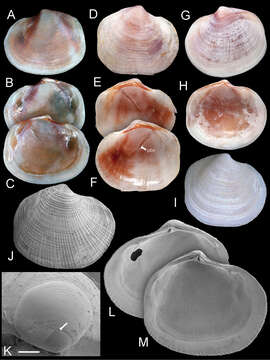
Figure 7.Ferrocina brunei sp. n. A–C Holotype NHMUK 20130122 Exterior of left valve and interior of right and left valves, L = 8.2 mm D–F Paratype NHMUK 20130123 exterior of right valve and interior of left and right valves, L = 8.4 mm. pbv trace of pallial blood vessel G–H Paratype NHMUK 20130123 exterior and interior of left valve, L = 8.9 mm I Exterior of right valve of white form NHMUK 20130123, L = 7.9 mm J SEM of right valve L = 6.0 mm K Protoconch, arrow at PI /PII junction. Scale bar = 50 µm L–M Interior of right and left valves L = 5.2 mm.
Included On The Following Pages:
- Life (creatures)
- Cellular (cellular organisms)
- Eukaryota (eukaryotes)
- Opisthokonta (opisthokonts)
- Metazoa (Animal)
- Bilateria
- Protostomia (protostomes)
- Spiralia (spiralians)
- Mollusca (molluscs)
- Bivalvia (mussels)
- Lucinida (Lucinoida)
- Lucinoidea
- Lucinidae
- Ferrocina
- Ferrocina brunei
This image is not featured in any collections.
Source Information
- license
- cc-by-3.0
- copyright
- John D. Taylor, Emily A. Glover
- bibliographic citation
- Taylor J, Glover E (2013) New lucinid bivalves from shallow and deeper water of the Indian and West Pacific Oceans (Mollusca, Bivalvia, Lucinidae) ZooKeys 326: 69–90
- original
- original media file
- visit source
- partner site
- Zookeys
- ID