Ventral infraciliature
Description:
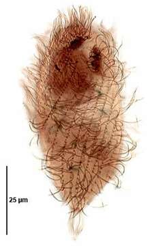
Infraciliature (ventral view) of the trichostomatid ciliate, Spirozona caudata (Kahl,1926). The cell is elongate and rounded anteriorly and tapers posteriorly to a narrow truncate cone. The cell is ellipsoid in cross section. The cytostome is located in the anterior �. Its right margin is curved and the left relatively straight. There are several oral polykinetids on the left and an undulating membrane on the right. The somatic ciliature is distinctive with a wide swath of closely spaced kineties spiraling from the right anterior to the posterior midline. A single spiral kinety of more densely packed kinetosomes bearing longer cilia originates to the right of the cytostome and spirals around the right side to the dorsum of the cell. There are 3 postoral kineties and one shorta paraoral kinetid on the left margin of the cytostome. More widely spaced kineties with less densely packed kinetids originate to the left of the cytostome and follow a less spiral course to the posterior end ventrally. The narrow truncate cone of the posterior end bears a circular row of kinetids. There is a small unciliated anterior apical area or "frontal plate". The spherical macronucleus and micronucleus are located in the anterior half. The single contractile vacuole is located at the posterior end. Collected from sapropelic bottom sediments from standing freshwater near Boise, Idaho November, 2004. Stained by the silver carbonate technique (see Foissner, W.Europ. J. Protistol.27,313-330;1991). Brightfield optics.
Included On The Following Pages:
- Life (creatures)
- Cellular (cellular organisms)
- Eukaryota (eukaryotes)
- SAR (Stramenopiles, Alveolates, Rhizaria)
- Alveolata (alveolates)
- Ciliophora (ciliates)
- Intramacronucleata
- Litostomatea
- Trichostomatia
- Spirozonidae
- Spirozona
- Spirozona caudata
This image is not featured in any collections.
Source Information
- license
- cc-by-nc
- author
- William Bourland
- provider
- micro*scope
- original
- original media file
- visit source
- partner site
- micro*scope
- ID