Infraciliature
Description:
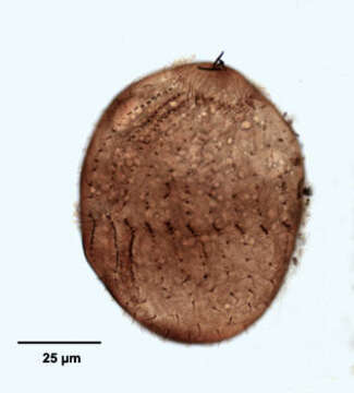
Right lateral view of the haptorid ciliate, Acropisthium mutabile (Perty, 1852). The cell body is ovoid to cylindrical. The posterior tapers to a short point. The fixation and staining process swells the cells. The anterior end forms a blunt snout with an apical cytostome. Short trichites support the cytopharynx (not seen here). There is a girdle of longer cilia just posterior to the bare anterior snout. There are 22 widely spaced uniform longitudinal somatic kineties. This individual is in the middle stage of division. The equatorial band of closely spaced kintosomes will form the circumoral ciliary girdle of the posterior daughter cell (opisthe). The anterior halves of three dorsal kineties are made up of clavate (short club-shaped) cilia forming a dorsal brush (seen well in this view). The dorsal brush of the opisthe is seen well here. Collected from freshwater pond near Boise, Idaho August 2004. This specimen is stained by a silver carbonate technique (see Foissner, W.Europ. J. Protistol.27,313-330;1991). Brightfield optics.
Included On The Following Pages:
- Life (creatures)
- Cellular (cellular organisms)
- Eukaryota (eukaryotes)
- SAR (Stramenopiles, Alveolates, Rhizaria)
- Alveolata (alveolates)
- Ciliophora (ciliates)
- Intramacronucleata
- Litostomatea
- Haptoria
- Haptorida
- Acropisthiidae
- Acropisthium
- Acropisthium mutabile
This image is not featured in any collections.
Source Information
- license
- cc-by-nc
- author
- William Bourland
- provider
- micro*scope
- original
- original media file
- visit source
- partner site
- micro*scope
- ID