Drawing
Description:
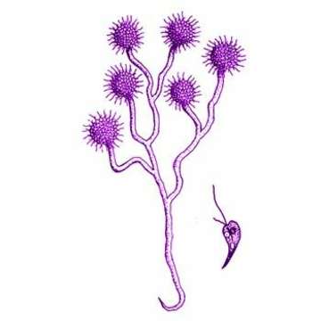
Anthophysa vegetans (Muller) Stein, 1878. Cells are wide at the anterior and at one side of the anterior end is a site where food is taken in, and the two flagella (one longer one shorter) insert to the side of this. The posterior tip of the cell extendS into a fine protoplasmic filament of variable length and many cells are usually united at their bases into spherical or hemispherical colonies, and these may either be free-swimming or attached to the substrate by means of a stalk which is coloured brown and is often branched. The stalk nearest the cells is usually narrower and transparent, becoming thickened distally by the deposition of iron and manganese compounds, bacteria are embedded in it throughout its length and can be seen clearly close to the colony.
Included On The Following Pages:
- Life (creatures)
- Cellular (cellular organisms)
- Eukaryota (eukaryotes)
- SAR (Stramenopiles, Alveolates, Rhizaria)
- Stramenopiles (heterokont)
- Ochrophyta (Ochrophyte)
- Chrysophyceae (golden algae)
- Chromulinales
- Chromulinaceae
- Anthophysa
- Anthophysa vegetans
- Oomycota (oomycetes)
- Chrysista
This image is not featured in any collections.
Source Information
- license
- cc-by-nc
- author
- D. J. Patterson.
- provider
- micro*scope
- original
- original media file
- visit source
- partner site
- micro*scope
- ID