portrait
Description:
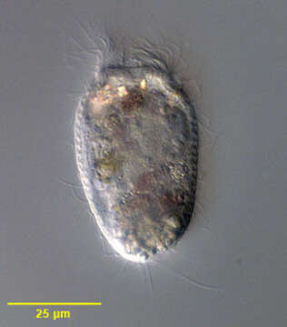
Portrait of free-swimming Vasicola ciliata (Tatem, 1869) that has vacated its lorica. Vasicola ciliata is a metacystid ciliate that produces a thin, transparent pseudochitinous vase-like lorica with shallow transverse corrugations (not seen in this image). In the lorica the cell body assumes a more globular shape. When swimming free the cell is more elongate. The circular cytostome is located in the center of the truncate anterior end. It opens into a cytopharynx supported by indistinct trichites. There are three concentric ciliary rings around the cytostome, the innermost with a single ciliary row, the middle with a double ciliary row and a third ring of four ciliary rows. The uniform longitudinal ciliary rows are formed of dikinetid kinetosomes the alignment which gives the appearance of transverse ciliary bands (paratenes). There is a sparse tuft of long caudal cilia (only one long eccentric posterior cilium is seen in the similar genus, Metacystis and another metacystid, Pelatractus, lacks caudal cilia). Multiple food vacuoles are visible in the cytoplasm. The central spherical macronucleus is seen in this image. The base of the lorica is partly filled with the expelled contents of defecation vacuoles. A single peripheral contractile vacuole is located in the posterior third of the cell (seen in this image on the viewer's left). A large, clear terminal vacuole may occur but is not seen in this image. The cell often vacates the lorica when disturbed. Vasicola ciliata is sapropelic and feeds on sulfur bacteria. Collected from stagnant freshwater sediment with strong smell of hydrogen sulfide near Boise, Idaho January 2004. DIC optics.
Included On The Following Pages:
- Life (creatures)
- Cellular (cellular organisms)
- Eukaryota (eukaryotes)
- SAR (Stramenopiles, Alveolates, Rhizaria)
- Alveolata (alveolates)
- Ciliophora (ciliates)
- Intramacronucleata
- Prostomatea
- Prostomatida
- Metacystidae
- Vasicola
- Vasicola ciliata
This image is not featured in any collections.
Source Information
- license
- cc-by-nc
- author
- William Bourland
- provider
- micro*scope
- original
- original media file
- visit source
- partner site
- micro*scope
- ID