-
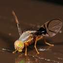
Yang Zai-Hua, Yu Jin-Yong, Yang Mao-Fa
Zookeys
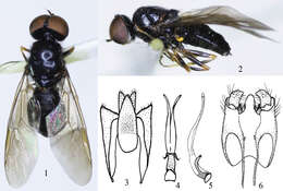
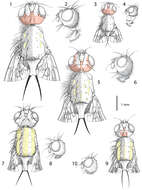
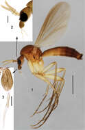
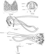
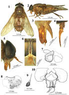
Figures 1–6.Oxycera ningxiaensis sp. n. holotype 1 Male, dorsal view 2 Male, lateral view 3 Proctiger, cerci and epandrium, ventral view 4–5 Aedeagal complex in dorsal and lateral view 6 Genital capsule, dorsal view.
-
Yang Zai-Hua, Yu Jin-Yong, Yang Mao-Fa
Zookeys
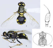
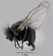
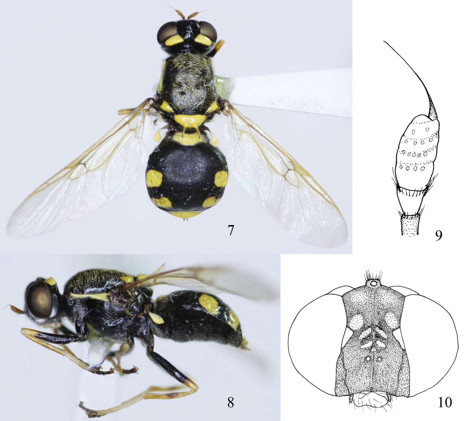
Figures 7–10.Oxycera rozkosnyi sp.n. holotype 7 Female, dorsal view 8 Female, lateral view 9 Antenna, inside 10 Head, frontal view.
-
Wayne N. Mathis, Tadeusz Zatwarnicki
Zookeys
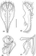
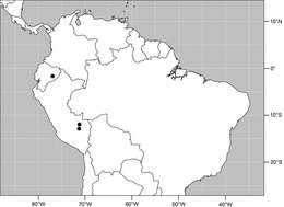
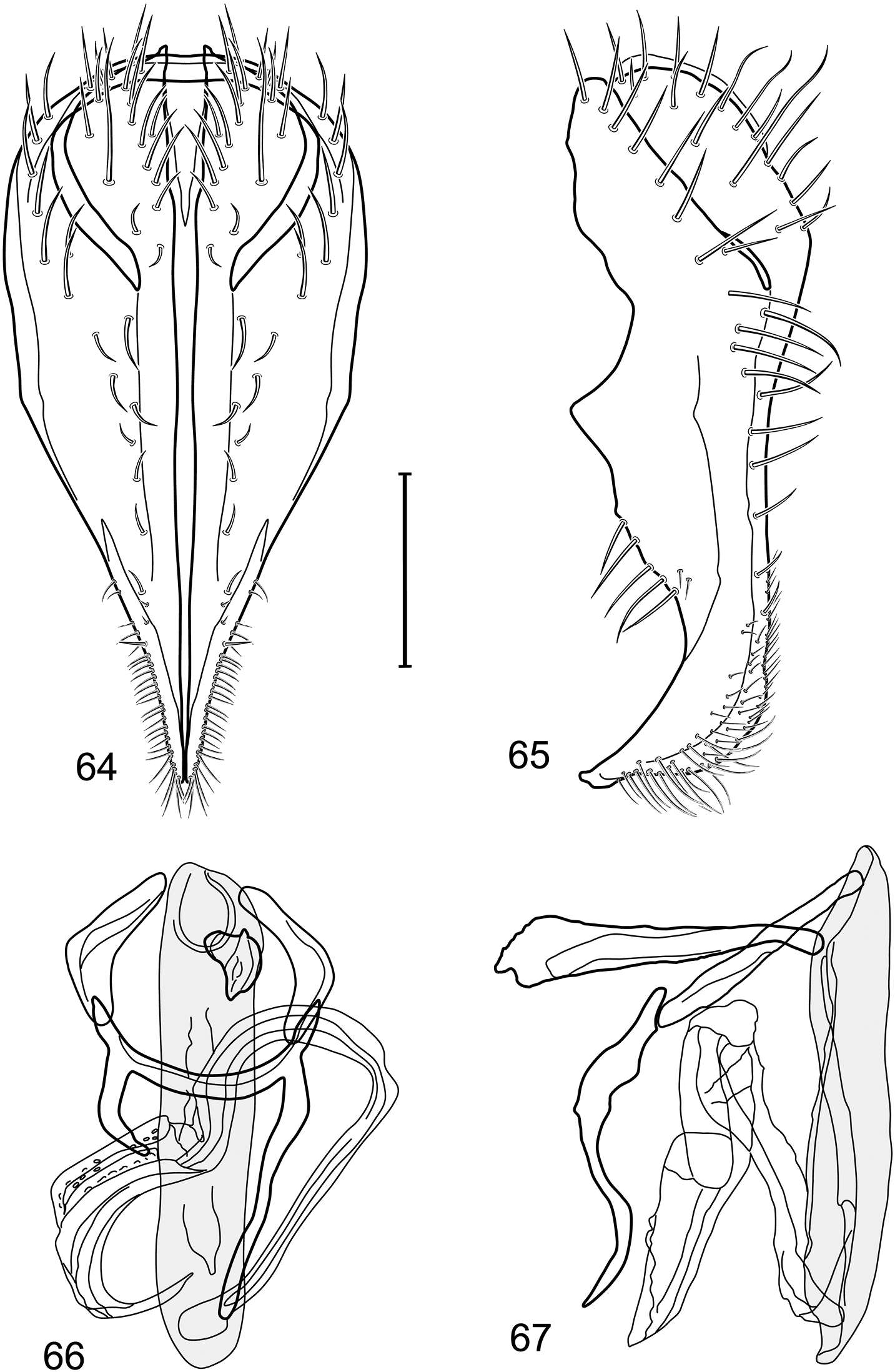
Figures 64–67.Illustration of Polytrichophora rostra sp. n. (male) (Ecuador. Napo: Pastaza) 64 epandrium and cerci, posterior view 65 same, lateral view 66 internal structures of male terminalia (aedeagus [shaded], phallapodeme, gonite, hypandrium), ventral view 67 same, lateral view. Scale bar = 0.1 mm.
-
Wayne N. Mathis, Tadeusz Zatwarnicki
Zookeys
Figure 68.Distribution of Polytrichophora rostra sp. n.
-
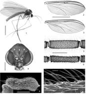
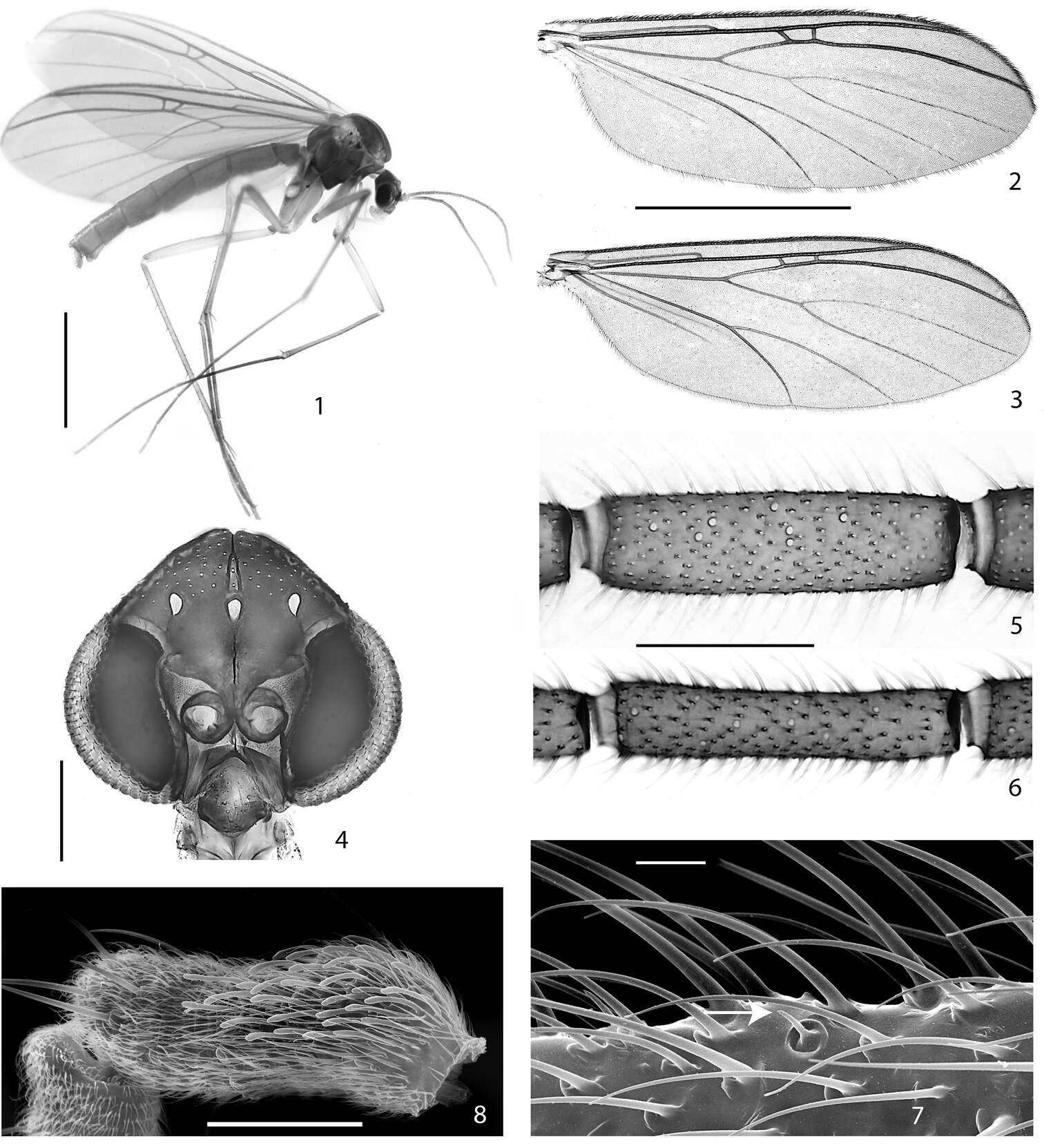
Figures 1–8.1–2, 4–5, 7–8. Acomopterella martinovskyi. 1 Female habitus 2 Male wing 4 Male head, frontal view 5 Flagellomere 4, male. Sensilla chaetica visible as pale spots. 7 Sensillum chaeticum (at arrowhead) on flagellomere 10, male 8 Sensilla on palpomere 3, male 3, 6 Acomopterella yoshiwaesp. n., male. 3 Wing 6 Flagellomere 4. Length of scale bar = 2 mm (for 1–3), 200 µm (for 4), 100 µm (for 5–6), 10 µm (for 7), 50 µm (for 8).
-
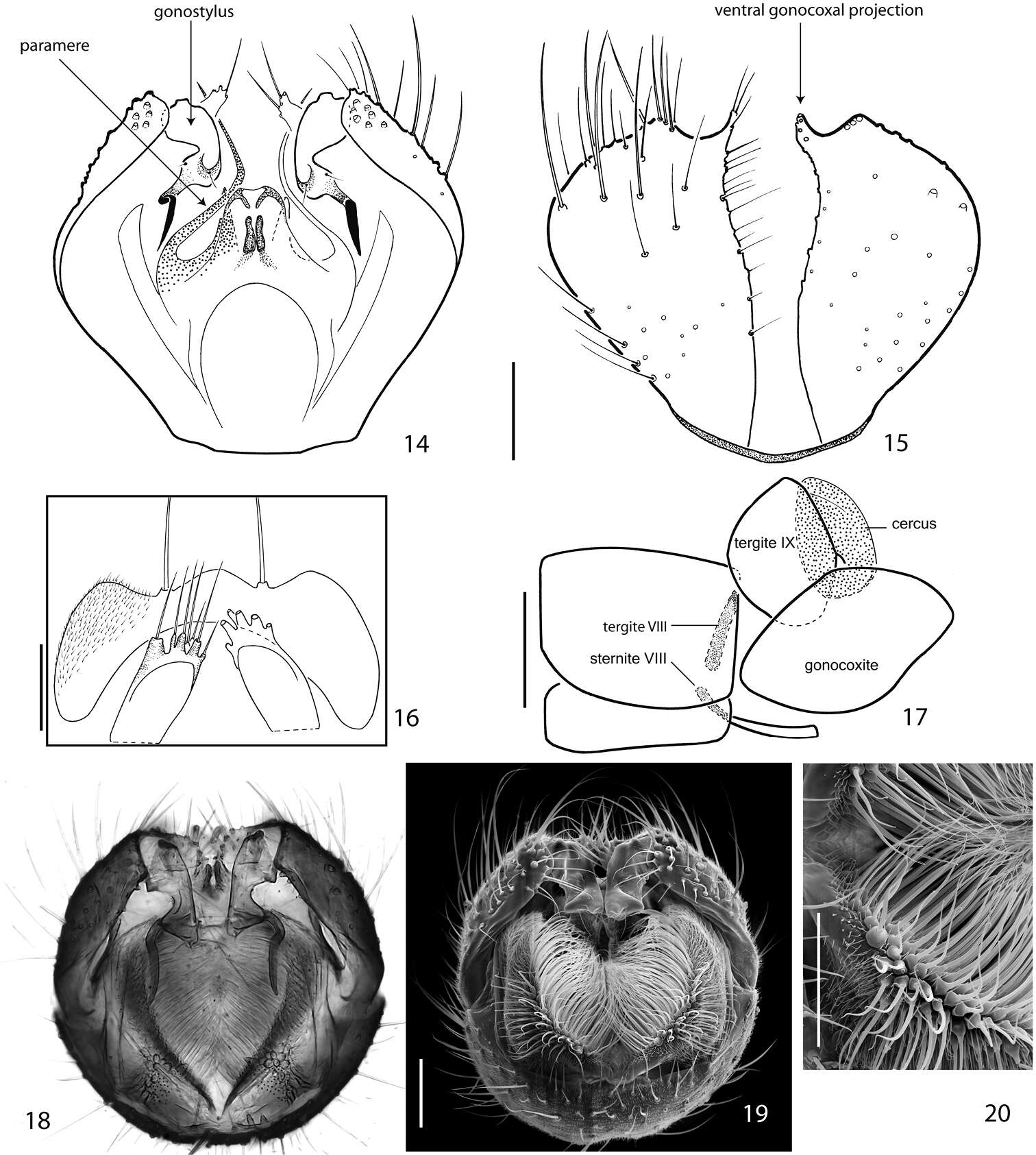
Figures 14–20.Terminalia of Acomopterella martinovskyi, male. 14 Terminalia in dorsal view, tergite IX removed 15 Gonocoxites in ventral view 16 Epiproct and hypoproct, dorsal view 17 Abdominal segments VII & VIII and terminalia in lateral view 18–19 Posterior view of terminalia 20 Cercal setae in detail. Length of scale bar = 100 µm (for 14–15, 18–19), 50 µm (for 16, 20), 0,5 mm (for 17).
-
Magdi S. El-Hawagry, Mohammed W. Khalil, Mostafa R. Sharaf, Hassan H. Fadl, Abdulrahman S. Aldawood
Zookeys
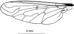
Figure 2.Wing of Anthrax alruqibi El-Hawagrysp. n.
-
Magdi S. El-Hawagry, Mohammed W. Khalil, Mostafa R. Sharaf, Hassan H. Fadl, Abdulrahman S. Aldawood
Zookeys
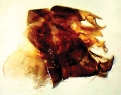
Figure 3.Male genitalia of Anthrax alruqibi El-Hawagrysp. n.
-
Maurizio Mei, Daniel Whitmore, Giuseppe Lo Giudice, Pierfilippo Cerretti
Zookeys
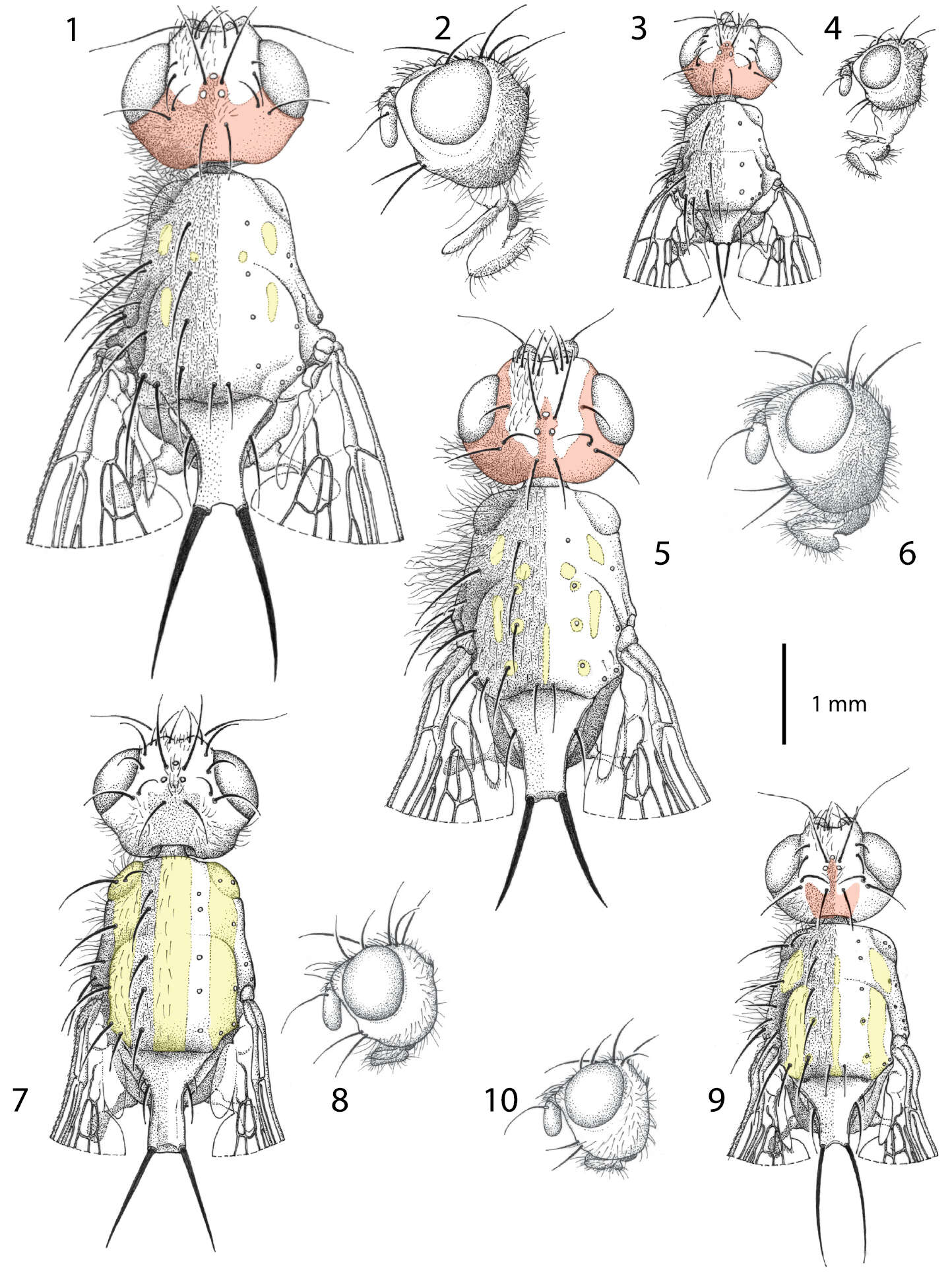
Figures 1−10.Males of Centrophlebomyia spp. 1, 3, 5, 7, 9 head and thorax in dorsal view 2, 4, 6, 8, 10 head in lateral view 1−4 Centrophlebomyia anthropophaga (Italy) 5−6 Centrophlebomyia furcata (Italy) 7−8 Centrophlebomyia grunini (Russian Far East) 9−10 Centrophlebomyia orientalis (India). In red the microtomentum pattern of head; in yellow the shiny, non microtomentose, pattern of thorax.
-
Maurizio Mei, Daniel Whitmore, Giuseppe Lo Giudice, Pierfilippo Cerretti
Zookeys
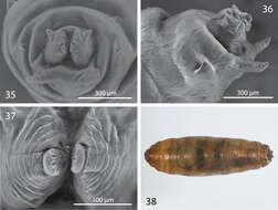
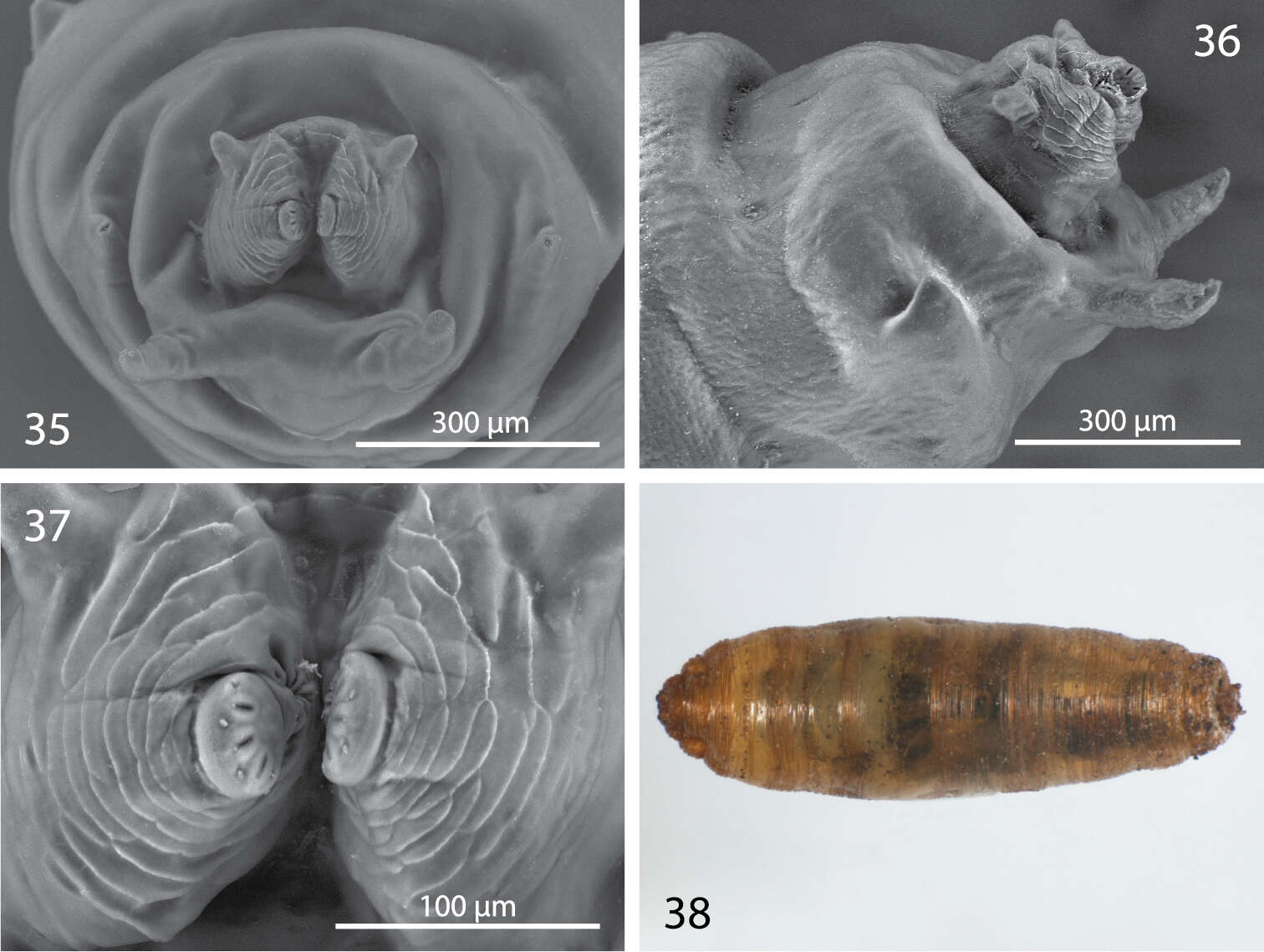
Figures 35−38.Third instar and puparium of Centrophlebomyia anthropophaga (Italy) 35 last segment bearing posterior spiracle in posterior view 36 last segment bearing posterior spiracle in lateral view 37 posterior spiracles in posterior view 38 puparium in dorsal view.
-
Xing Li, Xiao-long Lin, Xin-hua Wang
Zookeys
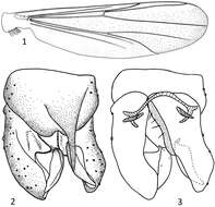
Figures 1–3.Parametriocnemus fortis sp. n., male. 1 wing 2 hypopygium(dorsal view) 3 hypopygium (ventral view).
-
Xing Li, Xiao-long Lin, Xin-hua Wang
Zookeys
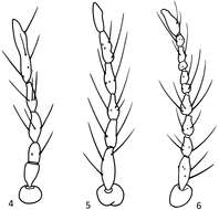
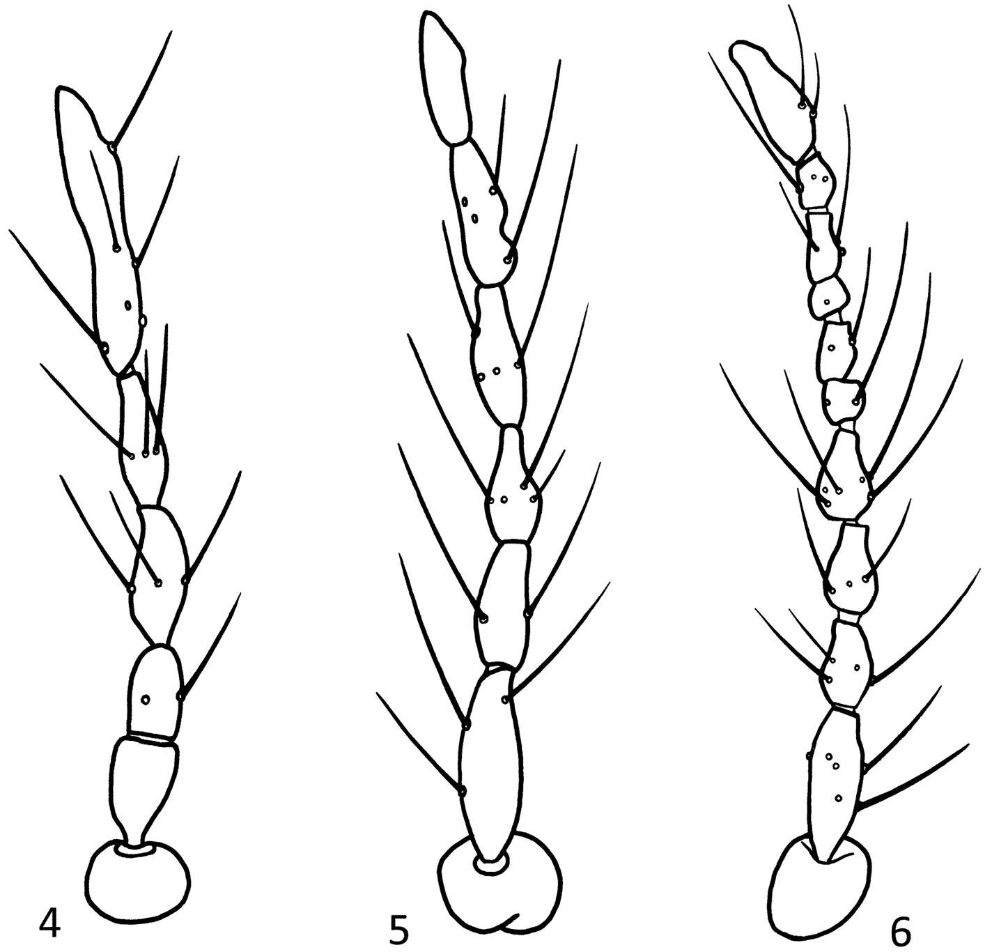
Figures 4–6.Parametriocnemus stylatus (Kieffer), intersex. 4 antenna, five segmented 5 antenna, six segmented; 6 antenna, ten segmented.
-
Mírian N. Morales, Gunilla Ståhls, Heikki Hippa
Zookeys
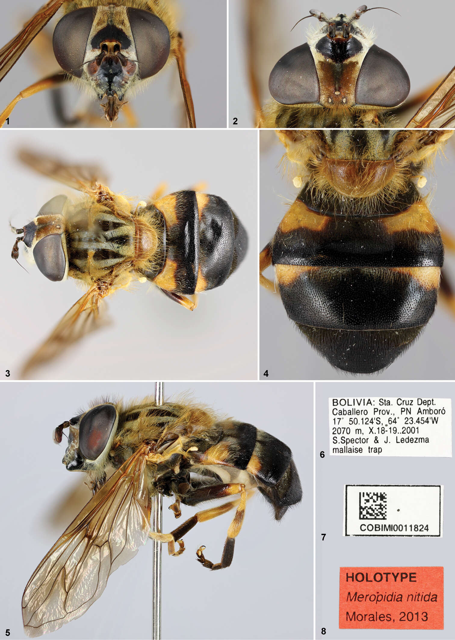
Figures 1–8.Meropidia nitida Morales, sp. n.,Holotype. 1 head, anterior view 2 head, dorsal view 3 habitus dorsal 4 abdomen, dorsal view 5 habitus lateral 6–8 labels.
-
Mírian N. Morales, Gunilla Ståhls, Heikki Hippa
Zookeys
Figures 9–15.Meropidia flavens Hippa & Ståhls, sp. n., Holotype 9 head, anterior view 10 head, lateral vie 11 habitus dorsa 12 habitus lateral 13–15 labels.
-
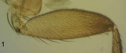
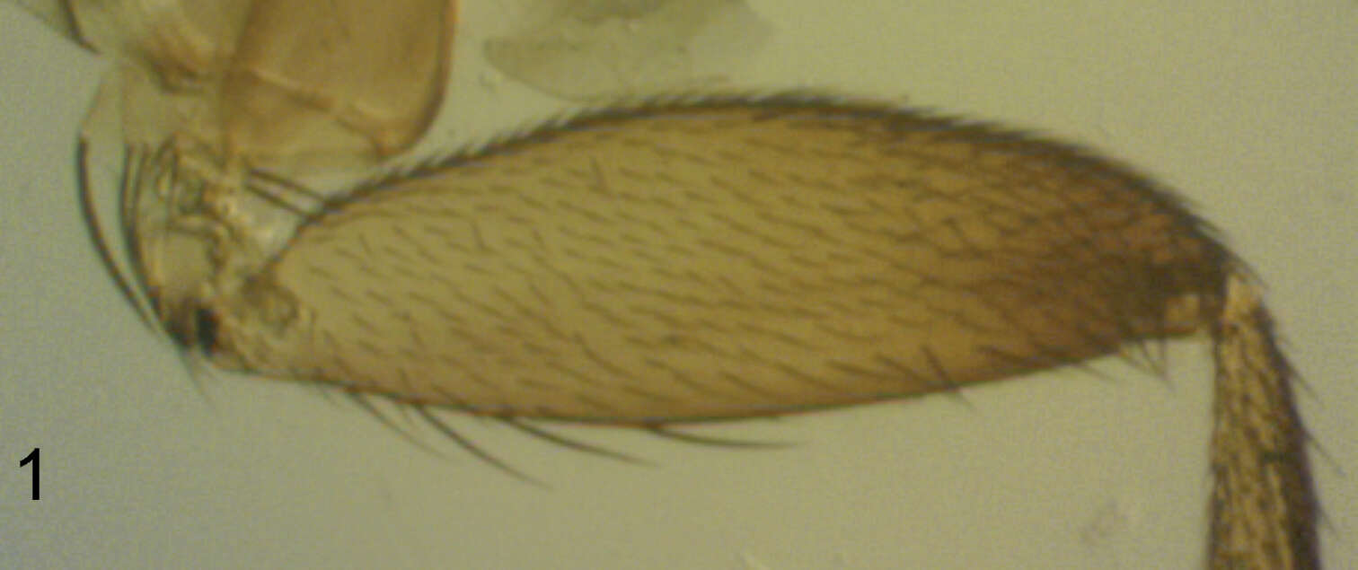
Figure 1.Megaselia elenae sp. n. male, anterior face of hind femur.
-
Olavi Kurina, Sarah Siqueira Oliveira
Zookeys
Figures 1–3.Cordyla australica sp. n. 1 male habitus 2 head with antennae and maxillary palpi, closer view 3 three apical segments of maxillary palpus. Scale bar = 1 mm (1), 0.2 mm (2) and 0.1 mm (3).
-
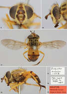
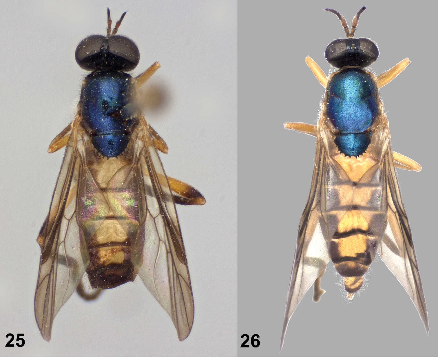
Figures 25–26.Dorsal views of Paraberismyia tzontehuitza Woodley. 25 Male (holotype) 26 Female (paratype).
-
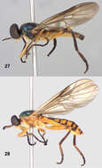
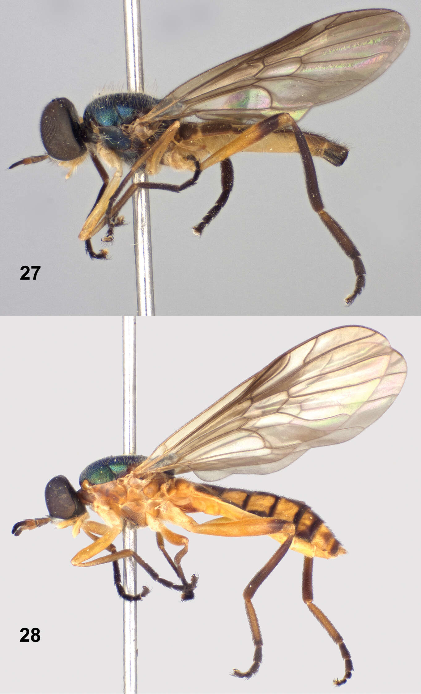
Figures 27–28.Left lateral views of Paraberismyia tzontehuitza Woodley. 27 Male (holotype) 28 Female (paratype).
-
Viviane Rodrigues de Sousa, Márcia Souto Couri
Zookeys
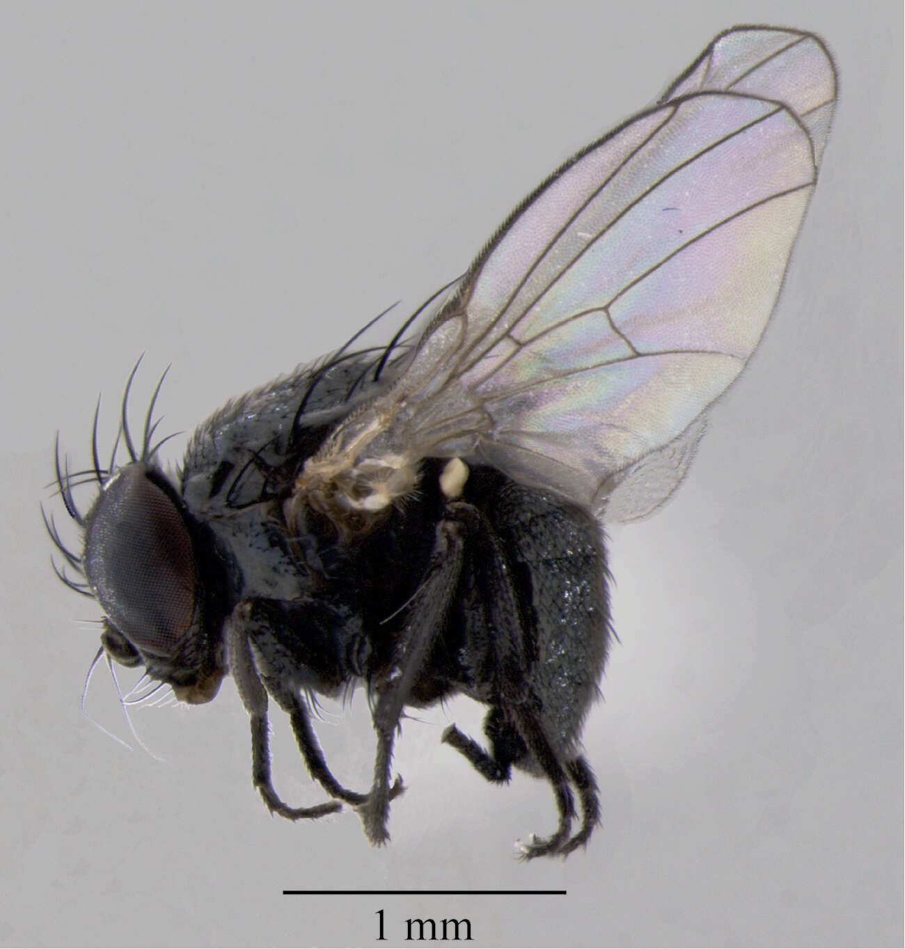
Figure 2.Japanagromyza inferna Spencer, male, in lateral view.
-
Viviane Rodrigues de Sousa, Márcia Souto Couri
Zookeys
Figures 3–7.Male terminalia of Japanagromyza inferna Spencer 3 sternite 5 4 epandrium, cercal plate and surstylus 5 hypandrium 6 phallapodeme, hypandrium, phallus 7 ejaculatory apodeme.
-
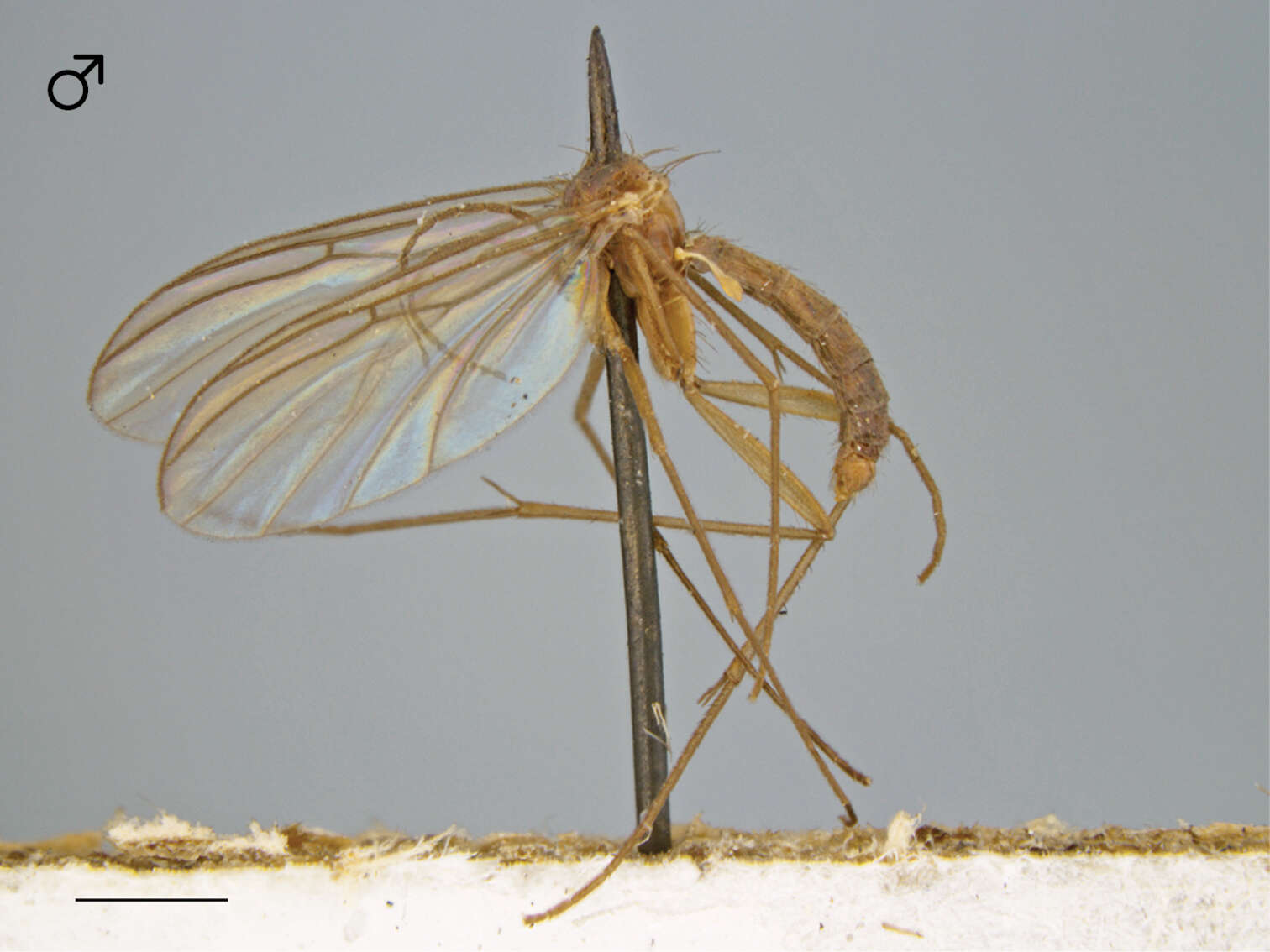
Figure 93.Ectrepesthoneura marceda (Sherman), habitus [male specimen # 598; 12K405]. Scale bar = 1 mm.
-
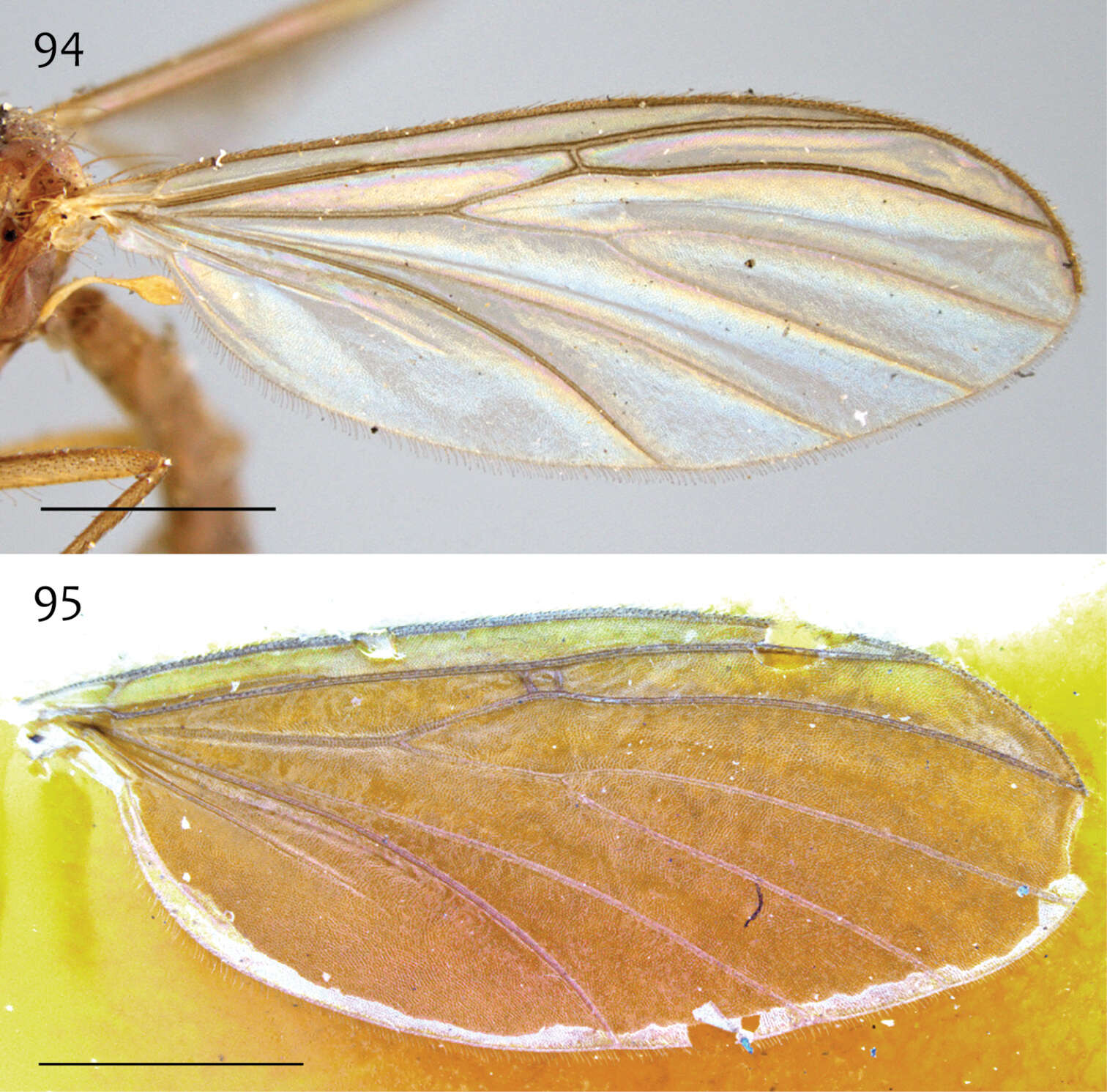
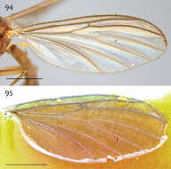
Figures 94–95.Ectrepesthoneura marceda (Sherman), wing 94 Specimen # 616; 13M406 95 Specimen # 329; 13M409. Scale bar = 1 mm.
-
Figures 1–10.Female Elephantotus tracuateuensis sp. n.: 1 Body in dorsal view 2 Body in lateral view 3 Palpus, labella and stilets 4 Antenna 5 Head in lateral view 6 Frons 7 Tergites 9, 10 and cerci 8 Sternite 8 and gonapophyses 9 Spermatheca 10 Genital furca.
-
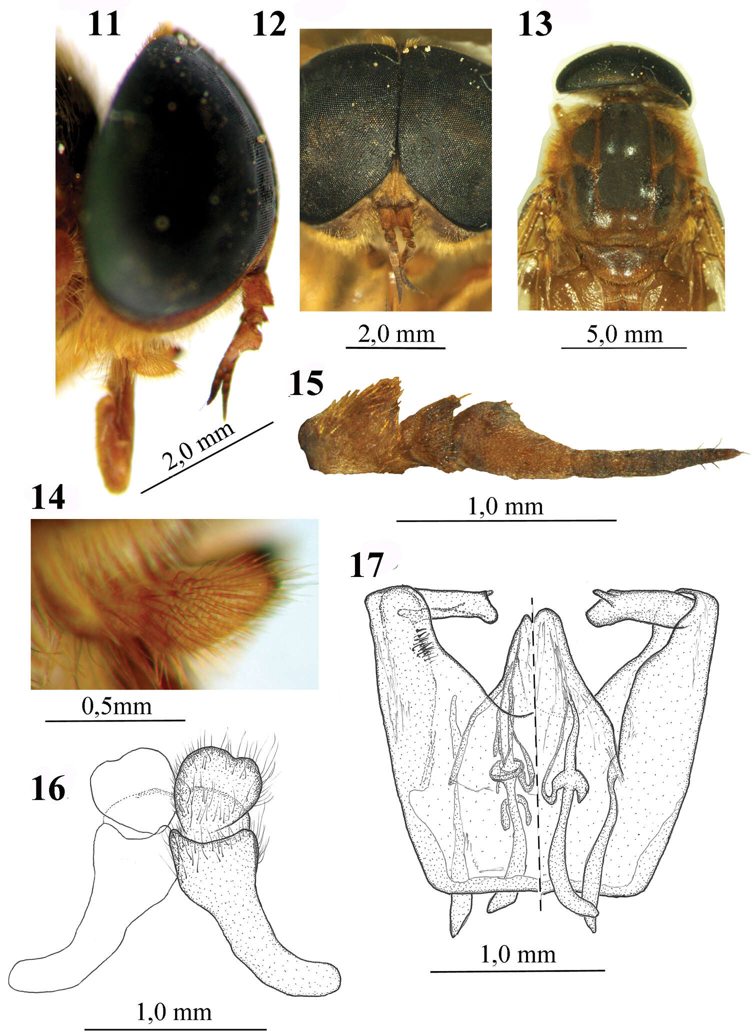
Figures 11–17.Male of Elephantotus tracuateuensis sp. n.: 11 Head in lateral view 12 Head in frontal view 13 Mesothorax and scutellum in dorsal view 14 Antenna 15 Palpus 16 Epandrium and cerci 17 Aedeagus.