fluorescence
الوصف:
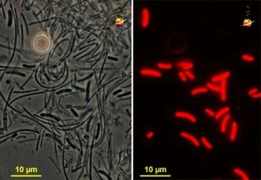
Synechococcus (sinm-eck-owe-cock-us), this pair of matched micrographs shows bacteria, mostly Synechococcus and Chloroflexus) from a mat sample. The phase contrast shot to the left shows the bacteria, the image to the right shows autofluorescence. Only the sausage-shaped Synechococcus exhibits autofluorescence. Phase contrast and fluorescence. Material provided by Mike Ferris from Mushroom Spring, a thermal site in Yellowstone National Park, photograph by Mike Ferris and David Patterson.
مشمول على الصفحات التالية:
- Life
- Cellular
- Bacteria
- Cyanobacteria (بكتيريا زرقاء)
- Synechococcales (متعاقبيات حبيبية)
- Synechococcaceae (متعاقبات حبيبية)
- Synechococcus (متعاقبة حبيبية)
- Terrabacteria
- Cyanobacteria/Melainabacteria group
هذه الصورة ليست واردة في أي مجموعات.
معلومات المصدر
- ترخيص
- cc-by-nc
- مؤلف
- David Patterson
- مقدم المحتوى
- micro*scope
- النص الأصلي
- ملف الوسائط الأصلي
- زيارة المصدر
- موقع الشريك
- micro*scope
- ID