-
This scanning electron micrograph (SEM) of an untreated water specimen extracted from a wild stream mainly used to control flooding during inclement weather, revealed the presence of unidentified organisms, which included bacteria, protozoa, and algae. In this particular image, a needle-shaped structure appeared to be caught up in an amorphous gelatinous biofilm, which though unidentified, appeared to be the green algae, Ankistrodesmus. See PHIL 11697 for a colorized version of this image.Created: 2009
-
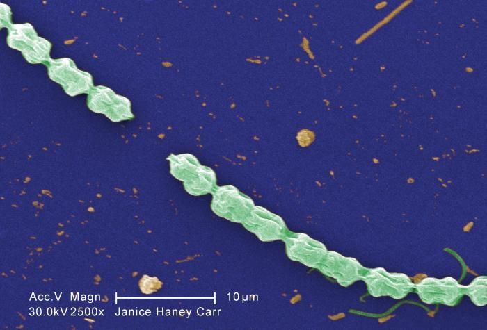
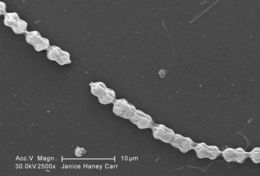
This scanning electron micrograph (SEM) of an untreated water specimen extracted from a wild stream mainly used to control flooding during inclement weather, revealed the presence of unidentified organisms, which included bacteria, protozoa, and algae. In this particular image, a filamentous chain of what appears to be a specie of green aglae known as Bulbochaete. Note what appeared to be one of this species characteristic hair cells on the right. The wrinkled appearance of this specimen may have been artifactual, brought on by having been processed prior to its examination under the electron microscope. See PHIL 11696 for a colorized version of this image.Created: 2009
-
This digitally-colorized scanning electron micrograph (SEM) of an untreated water specimen extracted from a wild stream mainly used to control flooding during inclement weather, revealed the presence of unidentified organisms, which included bacteria, protozoa, and algae. In this particular image, a filamentous chain of what appears to be a specie of green aglae known as Bulbochaete. Note what appeared to be one of this species characteristic hair cells on the right. The wrinkled appearance of this specimen may have been artifactual, brought on by having been processed prior to its examination under the electron microscope.Created: 2009
-
This scanning electron micrograph (SEM) of an untreated water specimen extracted from a wild stream mainly used to control flooding during inclement weather, revealed the presence of unidentified organisms, which included bacteria, protozoa, and algae. In this particular image, a filamentous chain of what appears to be a specie of green aglae known as Bulbochaete. Note what appeared to be one of this species characteristic hair cells on the right. The wrinkled appearance of this specimen may have been artifactual, brought on by having been processed prior to its examination under the electron microscope. See PHIL 11696 for a colorized version of this image.Created: 2009
-
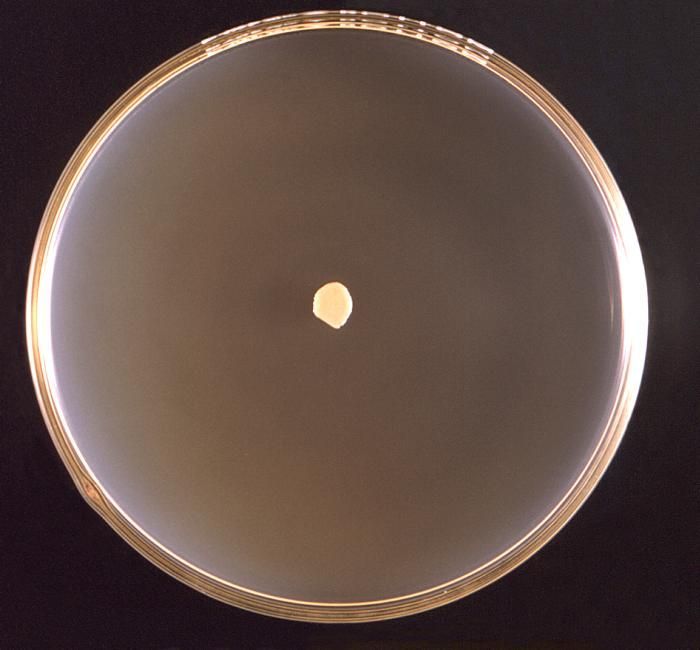
This 1971 image depicted a frontal view of a Petri dish culture in which a small colony of Prototheca wickerhamii algal organisms had been cultivated.Created: 1971
-
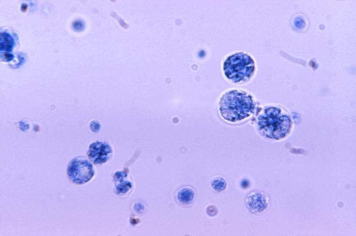
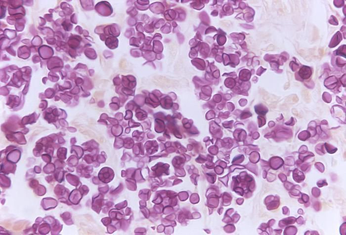
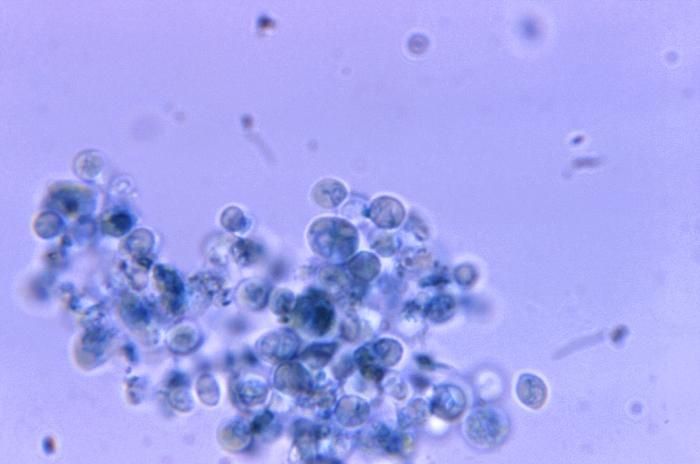
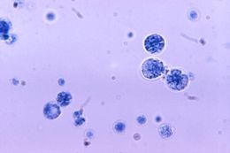
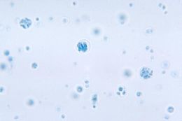
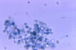
At a magnification of 1125X, this photomicrograph revealed the presence of a number of Prototheca wickerhamii algal organisms.Created: 1971
-
Under a magnification of 1125X, this photomicrograph revealed the presence of numbers of algal organisms, Prototheca wickerhamii, which were found within a tissue specimen. Though categorized taxonomically as an alga, it derives its sustenance as a saprophyte, consuming dead and decaying organic matter. This algal culture was prepared using a lactophenol cotton blue mount fixation technique.Under microscopic analysis, Prototheca spp. resemble a fungal organism, and can therefore, be mistaken when attempting to identify these algae.Similar to the members of the genus Chlorella, Prototheca spp. are heterotrophic, , which means these organisms require carbon in order to thrive, and obtains this nutrient through its consumption of organic substrates. This algal culture was prepared using a lactophenol cotton blue mount fixation technique.Created: 1972
-
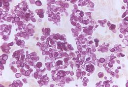
Under a magnification of 500X, this Gridley-stained photomicrograph revealed the presence of numbers of algal organisms, Prototheca wickerhamii, which were found within a specimen of deer tissue. Though categorized taxonomically as an alga, it derives its sustenance as a saprophyte, consuming dead and decaying organic matter.Under microscopic analysis, Prototheca spp. resemble a fungal organism, and can therefore, be mistaken when attempting to identify these algae.Similar to the members of the genus Chlorella, Prototheca spp. are heterotrophic, , which means these organisms require carbon in order to thrive, and obtains this nutrient through its consumption of organic substrates. This algal culture was prepared using a lactophenol cotton blue mount fixation technique.Created: 1972
-
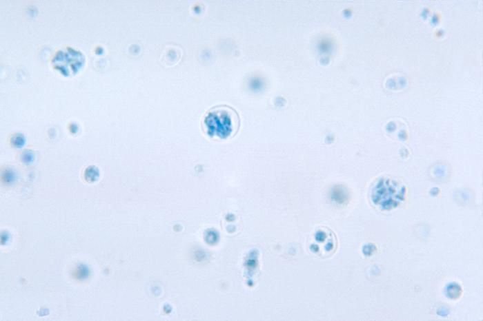
This photomicrograph depicts the presence of Prototheca wickerhamii in a case of protothecosis.Created: 1971
-
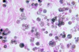
Note the histopathologic changes in protothecosis of the skin and mucous membrane of the nose due to P. wickerhamii.Created: 1973
-
This photomicrograph confirms the presence of Prototheca wickerhamii, an achlorophyllic algae.Created: 1972
-
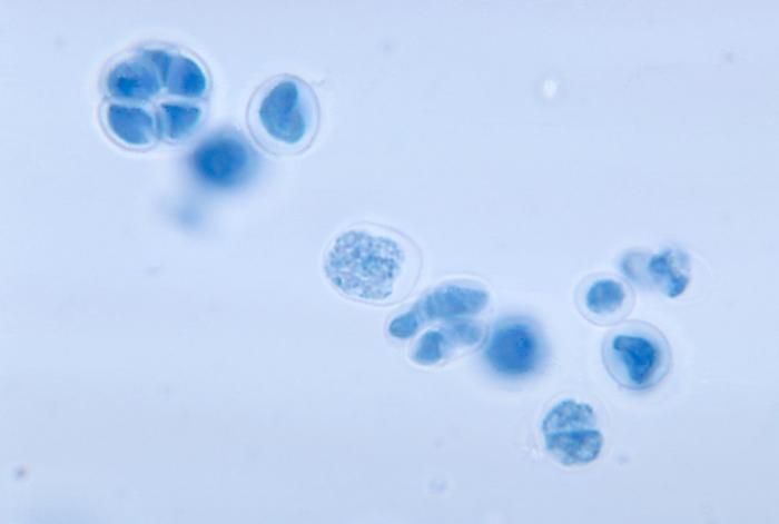
This photomicrograph depicts the presence of Prototheca wickerhamii using a lactophenol cotton blue mount technique.Created: 1972
-
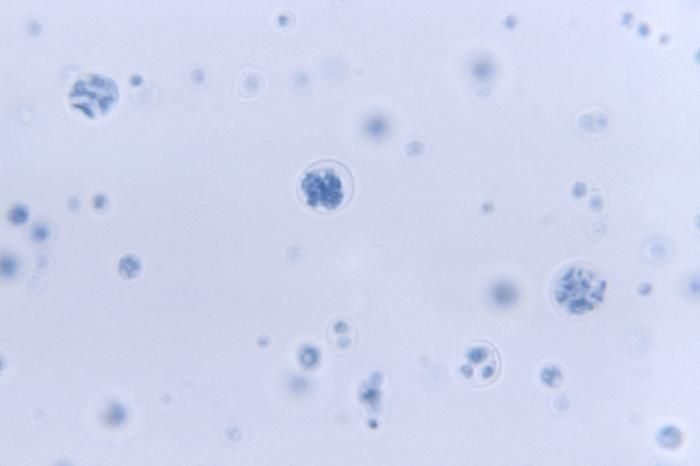
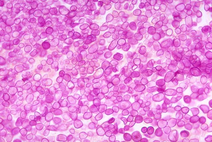
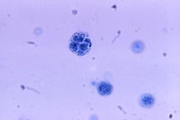
At a magnification of 1125X, this photomicrograph revealed the presence of a number of Prototheca zopfii algal organisms.Created: 1971
-
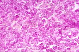
This photomicrograph depicts the histopathology associated with protothecosis in a dog due to Prototheca zopfii.Created: 1972
-
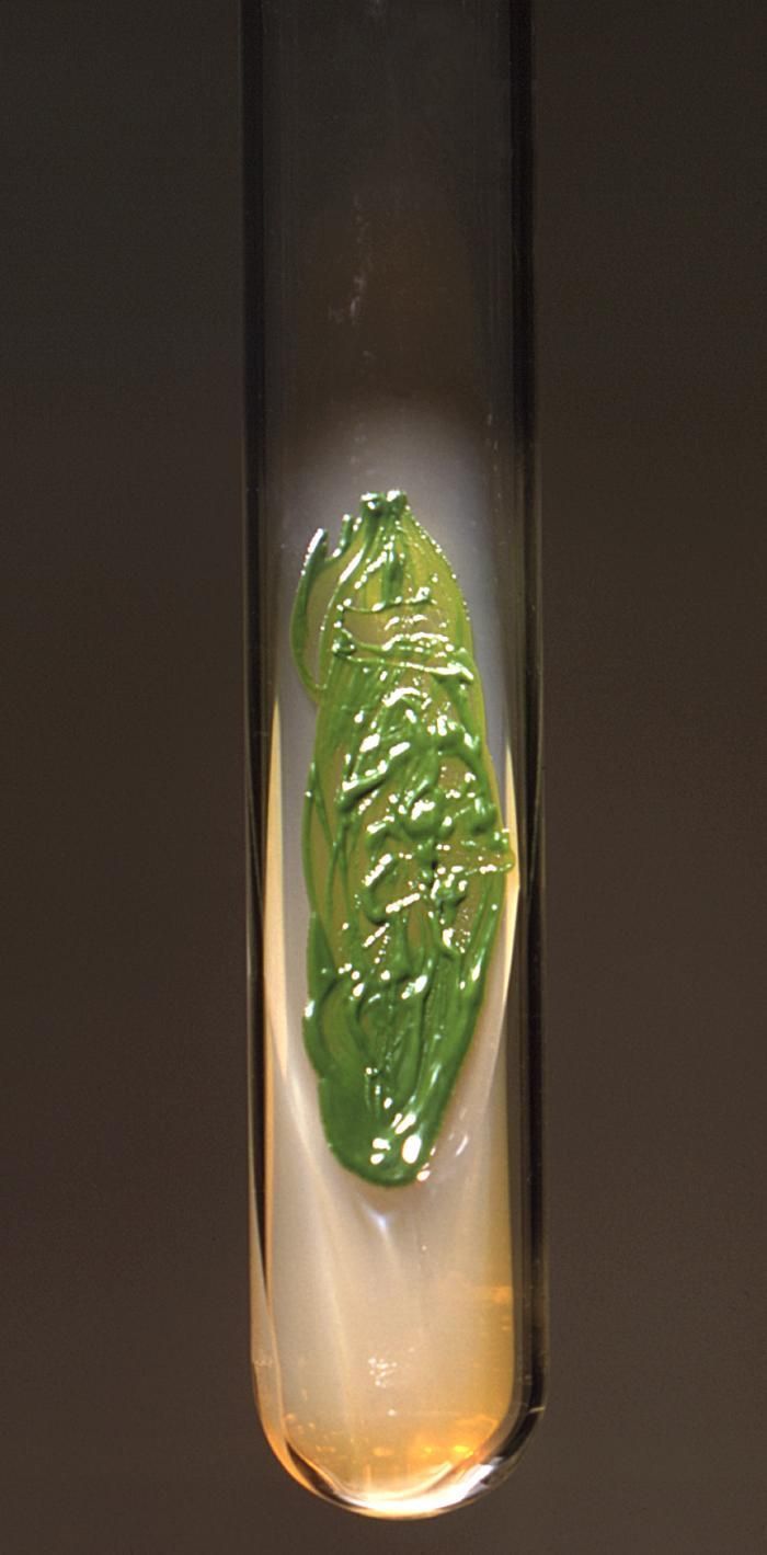
This 1971 image depicted a Sabourauds dextrose agar slant culture, which had cultivated a colony of Chlorella sp. algal organisms.Created: 1971
-
This 1971 image depicted a frontal view of a Petri dish culture in which a small coloney of Chlorella algal organisms had been cultivated.Created: 1971
-
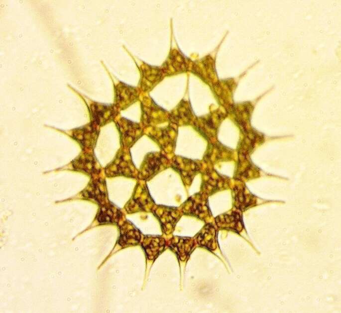
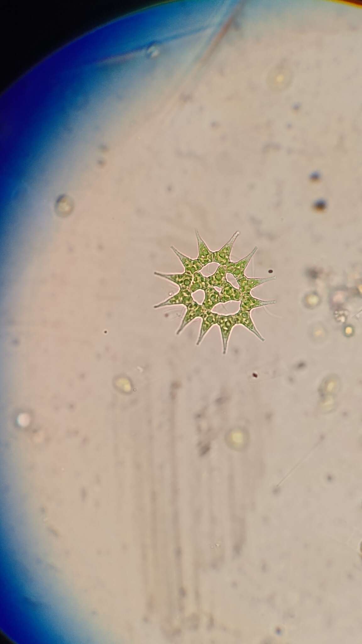
At a magnification of 1125X, this photomicrograph revealed the presence of a number of Chlorella sp. algal organisms.Created: 1971
-
-
-
-
-
-