-
The extrusomes are very difficult to see in vivo even with DIC. Exploded extrusomes are short and rod-shaped with rounded ends. The extrusomes are interposed between kinetids along the somatic kineties. Stained by the silver carbonate technique (see Foissner, W.Europ. J. Protistol.27:313-330;1991).Brightfield.
-
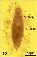
Fig 12: Horizontal ridges.
-
Posterior view of the infraciliature of Urotricha platystoma(STOKES,1886).The somatic kineties terminate in the posterior 1/4 of the cell (light blue arrowheads). The posterior end of the cell is unciliated (asterisk) except for the single long caudal cilium. the red arrowhead indicates the excentric pore of the contractile vacuole.Collected from the margin of a slow-moving outflow stream of a freshwater pond near Boise, Idaho.March 2007. Stained by the silver carbonate technique (Foissner,W. Europ. J. Protistol.27:313-330;1991).Brightfield.
-
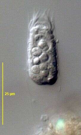
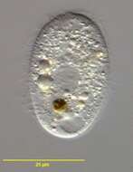
Collected from rewetted bottom sediments of a freshwater pond near Boise, Idaho, 2006. DIC.
-
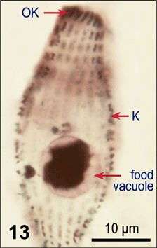
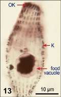
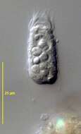
Fig 13 Protargol-stained cell, lateral view.
-
Urotricha platystoma (STOKES,1886).Collected from the margin of a slow-moving outflow stream of a freshwater pond near Boise, Idaho.March 2007. Phase contrast.
-
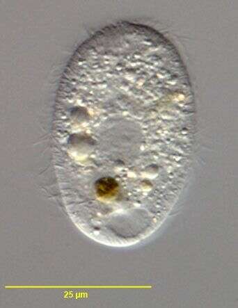
In vivo portrait of Metacystis borrori (ALADRO-LUBEL & MARTINEZ-MURILLO,2003). The cell body (10-35 X 10-18 um in vivo) is transversely annulated (4-6 rings). The somatic ciliature consists of 22-30 longitudinal kineties, and patterned as 5-7 transverse kineties. The circumoral kinety is composed of kinetosomes closely spaced. The macronucleus diam. about 3-7 pm. The lorica (18-61 X 11-26 um) with the posterior end round to conical or irregular and with mucoid filamemts. Collected from a commercial saltwater aquarium in Boise, Idaho.2004. Phase contrast.
-
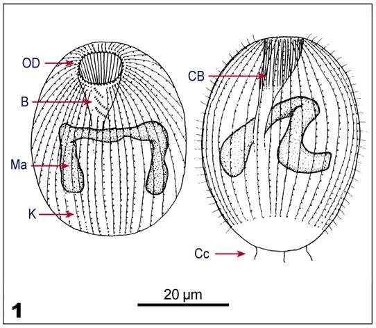
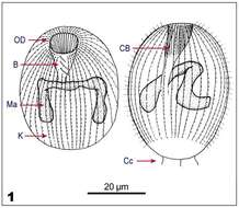
Fig 1 Line drawings of protargol stained cells, showing kineties, oral structures and nucleus.
-
Urotricha platystoma (STOKES,1886). The long caudal cilium is out of the focal plane in this image.Collected from the margin of a slow-moving outflow stream of a freshwater pond near Boise, Idaho.March 2007.DIC
-
In vivo portrait of Metacystis borrori (ALADRO-LUBEL & MARTINEZ-MURILLO,2003). The cell body (10-35 X 10-18 um in vivo) is transversely annulated (4-6 rings). The somatic ciliature consists of 22-30 longitudinal kineties, and patterned as 5-7 transverse kineties. The circumoral kinety is composed of kinetosomes closely spaced. The macronucleus diam. about 3-7 pm. The lorica (18-61 X 11-26 um) with the posterior end round to conical or irregular and with mucoid filamemts. Collected from a commercial saltwater aquarium in Boise, Idaho.2004. DIC.
-
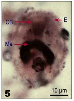
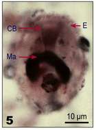
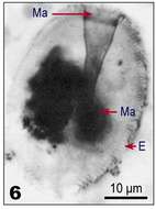
Fig 5: Protargol stains, Lateral view, showing macronucleus, cytopharyngeal basket, and extrusomes
-
Urotricha platystoma (STOKES,1886). Collected from the margin of a slow-moving outflow stream of a freshwater pond near Boise, Idaho.March 2007.DIC
-
In vivo portrait of Metacystis borrori (ALADRO-LUBEL & MARTINEZ-MURILLO,2003). The cell body (10-35 X 10-18 um in vivo) is transversely annulated (4-6 rings). The somatic ciliature consists of 22-30 longitudinal kineties, and patterned as 5-7 transverse kineties. The circumoral kinety is composed of kinetosomes closely spaced. The macronucleus diam. about 3-7 pm. The lorica (18-61 X 11-26 um) with the posterior end round to conical or irregular and with mucoid filamemts. Collected from a commercial saltwater aquarium in Boise, Idaho.2004. DIC.
-
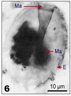
Fig 6: Protargol stains, Lateral view, showing macronucleus, cytopharyngeal basket, and extrusomes
-
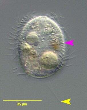
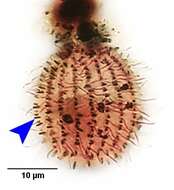
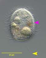
Urotricha platystoma (STOKES,1886). The yellow arrowhead indicates the single long caudal cilium.The pink arrowhead indicates the subcortical layer of fusiform extrusomes.Collected from the margin of a slow-moving outflow stream of a freshwater pond near Boise, Idaho.March 2007.DIC
-
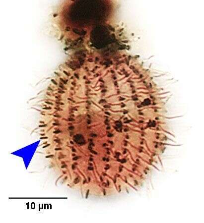
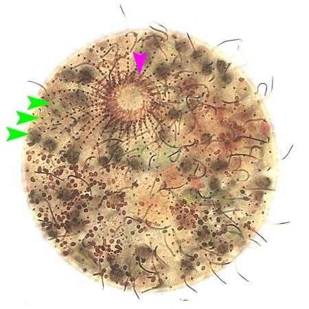
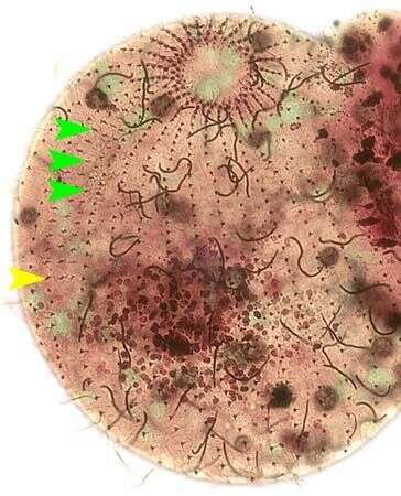
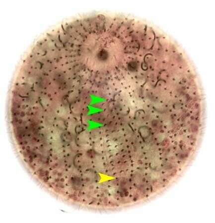
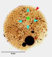
Infraciliature of Pelagothrix plancticola (FOISSNER,BERGER&SCHAUMBERG,1999).The three green arrowheads mark the three rows of dikinetids comprizing the adoral organelles or "dorsal brush".The rightmost adoral organelle is continuous posteriorly with a shortened somatic kinety.The pink arrowhead marks one of the dikinetids of the undulating membrane that borders the oral aperture.Specimen collected from an ephemeral freshwater puddle on the bank of the Boise river in Boise,Idaho.April 2007. Stained by the silver carbonate technique (Foissner,W. Europ. J. Protistol.27:313-330;1991).Brightfield.
-
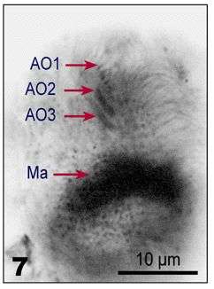
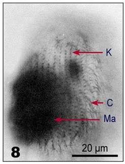
Fig 7: Protargol stains, Detail of the cortex, showing the three adoral rows of dikinetids (AO) of the brosse, the somatic kineties (K), and cilia (C).
-
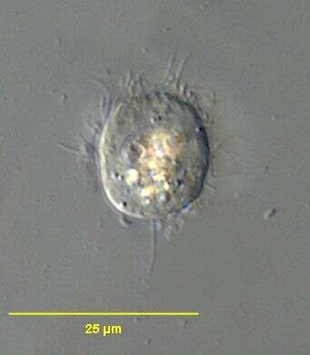
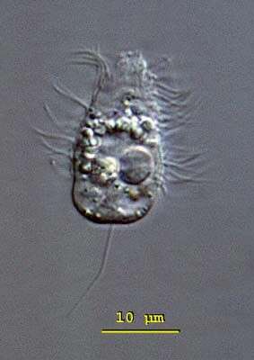
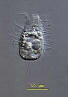
Urotricha (your-owe-trike-a) farcta: the anterior half of the body is conical and the posterior half trapeziform. The oral opening is at the anterior end of the cell. The contractile vacuole is located in the posterior end and the round macronucleus is in the middle of the body. The caudal cilium is slightly eccentric. This is a fast swimming cilate, the motion of which is interrupted by jumps. The species is mostly 15 - 30 microns long, this cell 22 microns. Differential interference contrast.
-
Infraciliature of Pelagothrix plancticola (FOISSNER,BERGER&SCHAUMBERG,1999).The three green arrowheads mark the three rows of dikinetids comprizing the adoral organelles or "dorsal brush".The rightmost adoral organelle is continuous posteriorly with a shortened somatic kinety (yellow arrowhead).Specimen collected from an ephemeral freshwater puddle on the bank of the Boise river in Boise,Idaho.April 2007. Stained by the silver carbonate technique (Foissner,W. Europ. J. Protistol.27:313-330;1991).Brightfield.
-
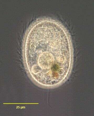
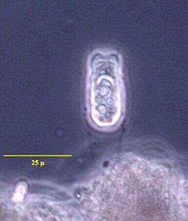
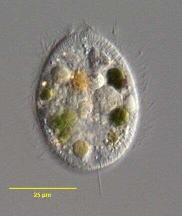
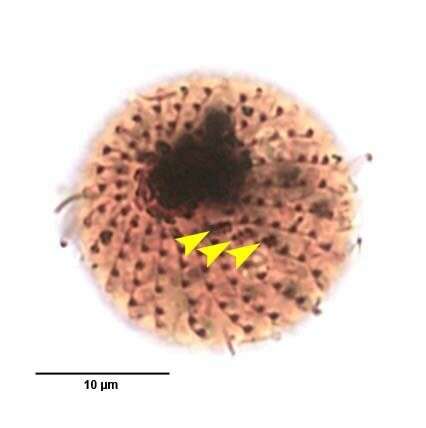
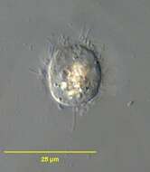
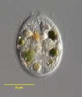
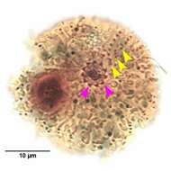
This ciliate from the Chesapeake Bay is a true omnivore having managed to ingest 3 pine pollen grains (the dark objects that resemble Mickey Mouse hats).
-
Fig 8: Protargol stains, Detail of the cortex, showing the three adoral rows of dikinetids (AO) of the brosse, the somatic kineties (K), and cilia (C).
-
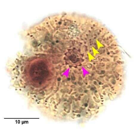
Oral infraciliature of Urotricha farcta (CLAPARÃDE&LACHMAN,1859). The pink arrowheads mark dikinetids of the undulating membrane (oral flaps).The yellow arrowheads mark the three minute adoral organelles. Collected from submerged dead leaves at the margin of a slow-flowing freshwater stream near Boise, Idaho.March 2007.Stained by the silver carbonate technique (Foissner,W. Europ. J. Protistol.27:313-330;1991).Brightfield.
-
Infraciliature of Pelagothrix plancticola (FOISSNER,BERGER&SCHAUMBERG,1999).The three green arrowheads mark the three rows of dikinetids comprizing the adoral organelles or "dorsal brush".The rightmost adoral organelle is continuous posteriorly with a shortened somatic kinety (yellow arrowhead).Specimen collected from an ephemeral freshwater puddle on the bank of the Boise river in Boise,Idaho.April 2007. Stained by the silver carbonate technique (Foissner,W. Europ. J. Protistol.27:313-330;1991).Brightfield.
-
Infraciliature of Urotricha farcta (CLAPARÃDE&LACHMAN,1859). The yellow arrowheads mark the three minute adoral organelles. Collected from submerged dead leaves at the margin of a slow-flowing freshwater stream near Boise, Idaho.March 2007.Stained by the silver carbonate technique (Foissner,W. Europ. J. Protistol.27:313-330;1991).Brightfield.