Image of Melloleitaoina uru Perafán & Pérez-Miles 2014
Description:
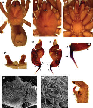
Figures 12–21.Melloleitaoina uru. 12 female, dorsal view 13–14 male holotype 13 cephalotorax 14 sternum, labium, maxillae and quelicerae 15 spermathecae 16–18 left palpal bulb 16 prolateral view 17 retrolateral view 18 detail of triangular tooth on embolus 19–20 coxa III 19 prolateral view 20 detail of spiniform setae 21 right tibial apophysis. Arrow indicates triangular tooth on embolus. Scale bars black = 1 mm.
Included On The Following Pages:
- Life (creatures)
- Cellular (cellular organisms)
- Eukaryota (eukaryotes)
- Opisthokonta (opisthokonts)
- Metazoa (Animal)
- Bilateria
- Protostomia (protostomes)
- Ecdysozoa (ecdysozoans)
- Arthropoda (arthropods)
- Chelicerata (chelicerates)
- Arachnida (arachnids)
- Araneae (spiders)
- Opisthothelae
- Mygalomorphae (mygalomorphs)
- Theraphosidae (tarantulas)
- Melloleitaoina
- Melloleitaoina uru
- Panarthropoda
This image is not featured in any collections.
Source Information
- license
- cc-by-3.0
- copyright
- Carlos Perafán, Fernando Pérez-Miles
- bibliographic citation
- Perafán C, Pérez-Miles F (2014) Three new species of Melloleitaoina Gerschman and Schiapelli, 1960 (Araneae, Mygalomorphae, Theraphosidae) from northern Argentina ZooKeys 404: 117–129
- original
- original media file
- visit source
- partner site
- Zookeys
- ID